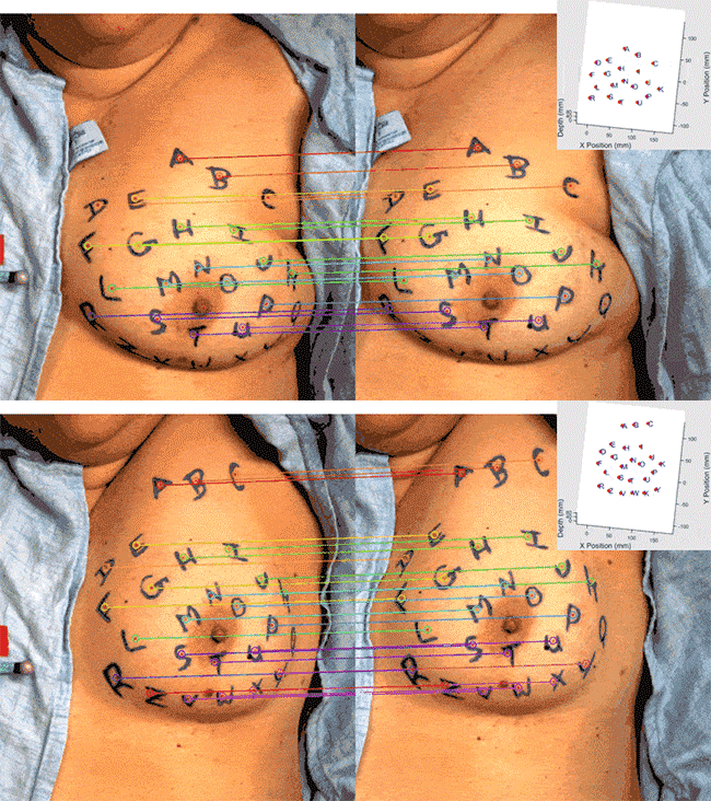
Acquiring data during surgery is often cumbersome. Existing methods to measure specific intraoperative points (fiducials) are generally time-consuming, imprecise, sparse, or some combination of these limitations. However, fast and accurate fiducial localization is necessary to provide seamless and trustworthy image-guided navigation. This work presents a contactless, automated fiducial acquisition method using stereo video of the operating field. The method provides reliable fiducial localization for an image guidance framework in breast conserving surgery.
On n=8 breasts from human subjects, the breast surface was measured throughout the full range of arm motion in a supine mock-surgical position. Using hand-drawn inked fiducials, adaptive thresholding, and KAZE feature matching, precise three-dimensional fiducial locations were detected and tracked through tool interference, partial and complete marker occlusions, significant displacements and nonrigid shape distortions.
Compared to digitization with a conventional optically tracked stylus, fiducials were automatically localized with 1.6 ± 0.5 mm accuracy and the two measurement methods did not significantly differ. The algorithm provided an average false discovery rate <0.1% with all cases’ rates below 0.2%. On average, 85.6 ± 5.9% of visible fiducials were automatically detected and tracked, and 99.1 ± 1.1% of frames provided only true positive fiducial measurements, which indicates the algorithm achieves a data stream that can be used for reliable on-line registration.
This method opens opportunities to move breast surgical guidance beyond initial single-shot rigid alignments and towards continuous correction and automatic updates to an image guidance system. This work-flow friendly data collection method provides highly accurate and precise three-dimensional surface data and is robust to occlusions, displacements, and most distortions of the skin surface. This demonstrates clinical utility of computer vision for monitoring soft tissue in the surgical field.

