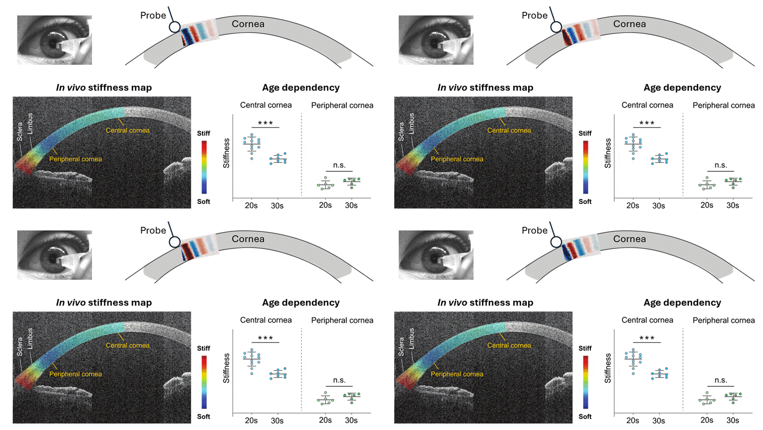
The mechanical properties of the cornea are pivotal in determining its response to external forces and have significant implications for vision care. Understanding corneal mechanics, particularly considering variabilities in stiffness, is essential for early detection of corneal ectasia diseases, accurate measurement of intraocular pressure (IOP), and optimizing outcomes in refractive surgeries. This study aims to develop a precise and reliable method for assessing the biomechanical properties of the human cornea in vivo.
We developed a high-frequency optical coherence elastography (OCE) technique using shear-like antisymmetric-mode Lamb waves at frequencies exceeding 10 kHz. We performed spatially-resolved OCE measurements in the sclera, limbus, and cornea on six subjects aged between 21 and 34. Furthermore, we obtained new in vivo data by applying the acoustoelastic theory to a corneal model accounting for IOP-induced tension, nonlinearities, anisotropy, and spatial variations of tissue stiffness.
Our study unveiled significant spatial variations in the shear modulus of the corneal stroma in healthy subjects for the first time. The central corneas displayed a mean shear modulus of 87 kPa, while a notable decrease to 44 kPa was observed in the corneal periphery. The central cornea’s shear modulus decreases with age with a slope of -19 +/- 8 kPa per decade, whereas the periphery showed non-significant age dependence. Additionally, we obtained wave displacement profiles that are consistent with the highly anisotropic nature of corneal tissues. The high-frequency OCE technique shows great potential for biomechanical assessment in clinical settings, offering valuable insights for refractive surgeries, diagnosing degenerative disorders, and assessing IOP.

