

Welcome to the BRAIN Special Issue! It consists of a group of 22 articles addressing the grand challenges in BRAIN: Brain Research through Advancing Innovative Neurotechnologies. These articles reflect a rich spectrum of BRAIN research on neurotechnologies for recording, imaging, interfacing and modulating the brain at multiple scales. The Issue was guest edited by: Fabio Babiloni, Walter Besio, Wei Chen, Lei Ding, Craig Henriquez, Pedro Irazoqui, Patrick Loughlin, Vasilis Marmarelis, Gernot Muller-Putz, Klaus-Robert Muller, Mohamad Sawan, Fan-Gang Zeng, and Babak Ziaie.

BEEG Source Imaging Enhances the Decoding of Complex Right-Hand Motor Imagery Tasks
Bradley J. Edelman, Bryan Baxter, Bin He, University of Minnesota, USA
Volume 63, Issue 1, Page: 4-14 Open Access
Brain-computer interfaces (BCIs) based on sensorimotor rhythms (SMRs) have achieved successful three-dimensional control using various motor imagination (MI) tasks. However, many control signals for state-of-the-art SMR BCIs are cognitively disconnected from action of the end effector.We extend previous EEG source imaging (ESI) work to decode four natural MI tasks of the right hand that may be utilized for realistic and intuitive BCI control. We report an increase of up to 18.6% for individual task classification, and an increase of 12.7% for the overall classification using the proposed ESI approach over the traditional sensor-based method.
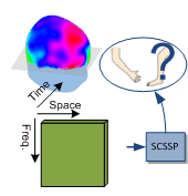
Separable Common Spatio-Spectral Patterns for Motor Imagery BCI Systems
Amirhossein S. Aghaei, Mohammad Shahin Mahanta, Konstantinos N. Plataniotis, University of Toronto, Canada
Volume 63, Issue 1, Page: 15-29
This paper addresses the problem of brain-computer interfacing using EEG signals obtained during motor imagery tasks, and focuses on spatio-spectral feature extraction in this context. Using a heteroscedastic matrix-variate Gaussian model, we introduce a novel theoretical approach for spatio-spectral generalization of the common spatial patterns (CSP) method. Compared to the alternative CSP generalizations, such as filterbank-CSP, the proposed algorithm has less computational cost and competent performance. Furthermore, it provides a theoretical measure for sorting the extracted features based on their discriminant power, which in turn eliminates the need for deployment of a separate feature selection step.
Decoding Brain States Based on Magnetoencephalography from Prespecified Cortical Regions
Jinyin Zhang, Xin Li, Stephen T. Foldes, Wei Wang, Jennifer L. Collinger, Douglas J. Weber, Anto Bagić, Carnegie Mellon University, & University of Pittsburgh, USA
Volume 63, Issue 1, Page: 30-42
We propose a novel region-of-interest-constrained discriminant analysis algorithm (RDA) for magnetoencephalography (MEG) decoding. RDA integrates linear classification and beamspace transformation into a unified framework so that brain states can be decoded by using signals from pre-specified cortical regions.The efficacy of RDA has been validated by the experimental results based on several human subjects. The proposed RDA tool offers a promising solution for multiple important applications. It may be used to train a target cortex during neurorehabilitation, study the functional roles of different cortical regions, or assist pre-surgical localization for patients with neurological disorders.
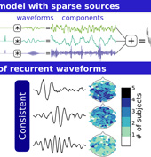
Learning Recurrent Waveforms within EEGs
Austin J. Brockmeier, Jose C. Principe, University of Liverpool, UK, University of Florida, USA
Volume 63, Issue 1, Page: 43-54
When experts analyze EEGs they look for phasic events of a particular frequency or waveform morphology. In this paper we explore a modeling approach that automatically learns the reoccurring waveforms within EEG traces. To summarize waveforms learned across electrodes and subjects we propose a cluster analysis protocol using shift-invariant k-means. The spatial amplitude patterns associated with a subset of the learned waveforms are shown to discriminate between motor imagery modalities.
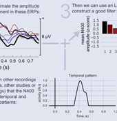
Single-trial ERP component analysis using a spatio-temporal LCMV beamformer
Marijn van Vliet, Nikolay Chumerin, Simon De Deyne, Jan Roel Wiersema, Wim Fias, Gerrit Storms, Marc M. Van Hulle, KU Leuven, Belgium
Volume 63, Issue 1, Page: 55-66
In the context of EEG analysis, beamformer filters are traditionally used for localization the source of some activity. In this paper, we show that they can be used for more than that. If the activation pattern of a specific ERP component (here, the N400) can be estimated (eg. by pooling data from all available recordings from all available subjects, perhaps even from multiple studies) these patterns can be combined with an estimation of the spatio-temporal covariance matrix of the current recording to design a filter that effectively isolates the desired ERP component on a single-trial basis.
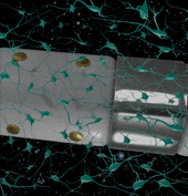
Combined single neuron unit activity and local field potential oscillations in a human visual recognition memory task
Kucewicz, M.T., Berry, B.M., Bower, M.R., Cimbalnik, J., Svehlik, V., Stead, S.M., Worrell, G.A., Mayo Clinic, USA
Volume 63, Issue 1, Page: 67-75
Current neurotechnologies typically probe a limited range of the vast scale of human brain electrophysiological activities, focusing either on the micro-electrode recordings of single neurons and their local assemblies or the macro-electrode field potential of large neural populations. This paper describes an analytical approach whereby high frequency oscillations and single neuron activity recorded from hybrid macro- and micro-electrodes during cognitive processing are both treated as point processes. In line with the multi-scale mapping goals of the BRAIN initiative the presented large-scale electrophysiology technique enables simultaneous sampling of individual neurons and their coordinated networks to study neuronal assemblies underlying human memory.
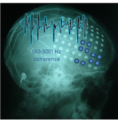
Spatial Coherence Profiles of Ictal High Frequency Oscillations Correspond to those of Interictal Low Frequency Oscillations in the ECoG of Epileptic Patients
Marija Cotic, Yotin Chinvarun, Martin del Campo, Peter L. Carlen, and Berj L. Bardakjian, University of Toronto, Canada; Hospital, Bangkok, Thailand; Toronto Western Hospital, Toronto, Canada
Volume 63, Issue 1, Page: 76-85
We examined the relationship between the spatiotemporal coherence patterns of slow and fast electrical oscillations in the brains of patients with extratemporal lobe epilepsy. We observed clusters of electrodes with strongly cohered slow oscillations-in between seizures-which typically exhibited strongly cohered fast oscillations-during seizures. This spatial overlap, of brain regions exhibiting both slow and fast coherence, highlighted seizure related regions of interest. As such, we propose these electrical rhythms as potential epilepsy biomarkers for extratemporal lobe seizures.

Analysis of Oscillatory Neural Activity in Series Network Models of Parkinson’s Disease During Deep Brain Stimulation
Clare M. Davidson, Annraoi M. de Paor, Hayriye Cagnan and Madeleine M. Lowery, University College Dublin, Ireland
Volume 63, Issue 1, Page: 86-95
Parkinson’s disease is a progressive, neurodegenerative disorder primarily affecting the motor system in humans. Recordings from the basal ganglia area of the brain show increased synchronous and oscillatory neural activity in Parkinson’s disease. Deep brain stimulation (DBS) suppresses this pathological activity, and is a successful way to control the symptoms of Parkinson’s disease for many people with the condition. We have developed a mathematical model that can be tuned to represent this pathological brain activity and its suppression with stimulation, providing a way to explore different DBS settings in silico.
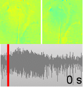
Dysfunction of Neurovascular/Metabolic Coupling in Chronic Focal Epilepsy
Yinchen Song, Rafael A. Torres, Sarahy Garcia, Yisel Frometa, Jihye Bae, Abhay Deshmukh, Wei-Chiang Lin, Ying Zheng, and Jorge J. Riera, Florida International University, USA & University of Reading, UK
Volume 63, Issue 1, Page: 96-110
In this article, we investigated the neurovascular/metabolic coupling in the epileptogenic cortices of rats with chronic focal epilepsy during ictal periods using a metabolically coupled balloon model, in comparison to normal rats undergoing forepaw stimulation. The results suggest that epileptogenic cortices have significantly larger hyperemic responses and a higher baseline of oxygen metabolism than normal cortices, which could positively impact on the interpretation of neuroimaging data in clinical practice. This is also the first study capturing the spatiotemporal electrophysiological and hemodynamic signatures from in vivo brains of rats with chronic focal epilepsy during ictal periods.
Chronic in vivo evaluation of PEDOT/CNT for stable neural recordings
Takashi D.Y. Kozai, Kasey Catt, Zhanhong Du, Kyounghwan Na, Onnop Srivannavit, Razi-ul M. Haque, John Seymour, Kensall D Wise, Euisik Yoon, X. Tracy Cui, University of Pittsburgh, University of Michigan, ePack, Inc, Structured Microsystems LLC, USA
Volume 63, Issue 1, Page: 111-119
Sub-cellular sized chronically implanted recording electrodes have demonstrated significantly improved tissue interface and single-unit recording performance. In order to overcome the high impedance barrier from ultrasmall recording sites, low impedance electrode coatings have been of significant interest. We report that a coating made of a new blend of PEDOT doped with carboxyl functionalized multi-walled carbon nanotubes demonstrated significant (p<0.05) long-term recording improvement over the traditional PEDOT/PSS formula on silicon lattice electrode arrays.
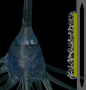
Close-Packed Silicon Microelectrodes for Scalable Spatially Oversampled Neural Recording
Jorg Scholvin, Justin P. Kinney, Jacob G. Bernstein, Caroline Moore-Kochlacs, Nancy Kopell, Clifton G. Fonstad, Edward S. Boyden, Massachusetts Institute of Technology & Boston University, USA
Volume 63, Issue 1, Page: 120-130 Open Access
Neural recording electrodes are important tools for understanding neural codes and brain dynamics. Here we present the design and implementation of close-packed silicon microelectrodes, to enable scalable spatially oversampled recording of neural activity, which facilitates data analysis. Our probes are fabricated in a dense array of recording sites connected to submicron dimension wiring. We demonstrate an implementation of a probe with 9 x 9 μm recording sites at 11 μm pitch and perform neural recordings with such probes in the live mammalian brain, illustrating the spatial oversampling potential of closely packed electrode sites.
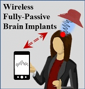
A Wireless Fully-Passive Neural Recording Device for Unobtrusive Neuropotential Monitoring
Asimina Kiourti, Cedric W.L. Lee, Junseok Chae, and John L. Volakis, The Ohio State University, Arizona State University, USA
Volume 63, Issue 1, Page: 131-137
We report a wireless and fully-passive brain implant, i.e. no wire, no battery, no rectifier, no energy harvester. The implant is capable of inconspicuous acquisition of very low-level neuropotentials down to 63µVpp in the frequency of 100 Hz – 5 kHz. The proposed implant improved the sensitivity by a factor of 8 over prior work. Such high sensitivity with sufficient bandwidth implies reading of most neuropotentials generated by the human brain. This capability of the neurosensor provides for transformational health-status monitoring possibilities for epilepsy, prosthetics control, Alzheimer’s disease, and many neurological disorders.
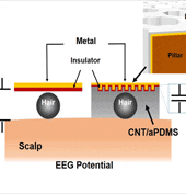
Self-adhesive and Capacitive Carbon Nanotube-based Electrode to Record Electroencephalograph Signals from the Hairy Scalp
Seung Min Lee, Jeong Hun Kim, Cheolsoo Park, Ji-Young Hwang, Joung Sook Hong, Kwang Ho Lee, Sang Hoon Lee, Gwangwoon University, Soongsil University, Kangwon National University, Korea University, South Korea
Volume 63, Issue 1, Page: 138-147
For brain-computer-interface, it is essential to record continuous electroencephalograph (EEG) with high signal quality. In this paper, we introduced a novel EEG electrode that is composed of uniformly distributed carbon nanotube (CNT) embedded within adhesive polydimethylsiloxane (aPDMS). This compliant conductive electrode allows it to conform to the shape of contacting surface. Thus, EEG could be measured accurately even with hairs on the contacting surface. With the capacitive coupling in the interface between metal and CNT/ aPDMS composite, the insulation layer is controllable. Lastly, to amend the EEG quality, pillars were implemented and Parylene C layer was deposited as an insulation layer.
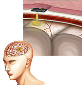
A Novel Lead Design for Modulation and Sensing of Deep Brain Structures
Allison T. Connolly,Rio J. Vetter, Jamille F. Hetke, Benjamin A. Teplitzky, Daryl R. Kipke, David S. Pellinen, David J. Anderson, Kenneth B. Baker, Jerrold L. Vitek, and Matthew D. Johnson, University of Minnesota, NeuroNexus, USA
Volume 63, Issue 1, Page: 148-157
This work aims to develop and characterize the functionality of a novel thin-film probe technology with a higher density of electrode contacts than are currently available with commercial deep brain stimulation (DBS) lead technology. Such technology has potential to enhance the spatial precision of DBS and enable a more robust approach to sensing local field potential activity in the context of adaptive DBS strategies. These 3D DBS arrays provide an enabling tool for future studies that seek to monitor and modulate deep brain activity through chronically implanted leads and has potential to enable sculpting DBS current flow and sensing biomarkers of disease and therapy.
Computational Study on the Thermal Effects of Implantable Magnetic Stimulation Based on Planar Coils
Hee Jin Park, Jae Hun Seol, Jeonghun Ku, Sohee Kim, Gwangju Institute of Science and Technology, and Keimyung University, Korea
Volume 63, Issue 1, Page: 158-167
For magnetic stimulation coils to be implantable in the brain, the temperature increase induced in the brain needs to be minimized as it is thought to be the primary source of adverse effects. We conducted computational simulations on the thermal effects of implantable magnetic stimulation using finite element analysis, by varying geometric parameters of planar spiral coils and repetitive stimulation pulse patterns. It is revealed that the temperature increase can be controlled by a careful design of coils and the most critical parameter is the outer diameter. The coil diameter greater than 8 mm is required to induce an allowable temperature rise of less than 1°C.
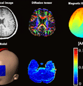
Current Density Imaging during Transcranial Direct Current Stimulation using DT-MRI and MREIT: Algorithm Development and Numerical Simulation
Oh In Kwon, Saurav Z. K. Sajib, Igor Sersa, Tong In Oh, Woo Chul Jeong, Hyung Joong Kim, and Eung Je Woo
Volume 63, Issue 1, Page: 168-175
We developed a new MR-based current density imaging technique to monitor the current flow inside the brain during tDCS (transcranial direct current stimulation). We validated the current density imaging method using a realistic three-dimensional human head model, measured DT-MRI images, and simulated magnetic flux density data. We found that the method can reliably and quantitatively visualize internal current density distributions during tDCS treatments.
Numeric Investigation of Brain Tumor Influence on the Current Distributions during Transcranial Direct Current Stimulation
Bo Song, Peng Wen, Tony Ahfock and Yan Li, University of Southern Queensland, Australia
Volume 63, Issue 1, Page: 176-187
This work is supposed to be the first numerical study of tDCS applications on the patients with brain tumors and our stimulation result showed that it should be safe to treat the neuropsychiatric conditions and cancer pain caused by the brain tumor using tDCS, though considerable global and local changes of the current distributions are induced by the present of a brain tumor. These findings should be helpful for tDCS researchers and clinical doctors to understand the current distributions of the brain with tumors and treat patients with anatomical distortions.
Development and Validation of a High Fidelity Finite Element Model of Monopolar Stimulation in the Implanted Guinea Pig Cochlea
Paul Wong, Shefin George, Phillip Tran, Andrian Sue, Paul Carter, and Qing Li, The University of Sydney, The Bionics Institute, Cochlear Limited, Australia
Volume 63, Issue 1, Page: 188-198
In this study, a highly detailed electroanatomical model of the cochlea was developed for investigating current flow in the implanted ear. Spatial representations of the current paths and corresponding neural responses revealed the underlying physical response to electrical stimulation. The model showed a strong correlation with independently obtained in vivo data, building confidence for using the model in future investigations.
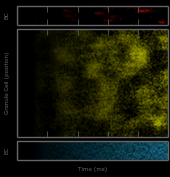
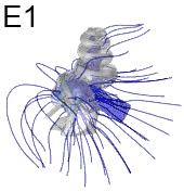
A Million-Plus Neuron Model of the Hippocampal Dentate Gyrus: Critical Role for Topography in Determining Spatio-Temporal Network Dynamics
Phillip J. Hendrickson, Gene J. Yu, Dong Song, and Theodore W. Berger, University of Southern California, USA
Volume 63, Issue 1, Page: 199-209
This work describes a million-plus granule cell compartmental model of the rat hippocampal dentate gyrus, including excitatory, perforant path input from the entorhinal cortex, and inhibitory input from dentate interneurons. Perforant path input was modeled based on axonal transport studies documenting the topographical organization of projections from subregions of the entorhinal cortex. Results showed that when entorhinal cortical neurons maintained Poisson random firing, dentate granule cells throughout the network expressed a robust pattern of structured spiking best described as spatio-temporal “clustering.” Findings conclusively identified topographical organization of inputs as the key element responsible for generating these clusters.
Spiking Neural Network with Distributed Plasticity Reproduces Cerebellar Learning in Eye Blink Conditioning Paradigms
Alberto Antonietti, Claudia Casellato, Jesús A. Garrido, Niceto R. Luque, Francisco Naveros, Eduardo Ros, Egidio D’Angelo, and Alessandra Pedrocchi, Politecnico di Milano, University of Granada, University of Pavia, Italy
Volume 63, Issue 1, Page: 210-219
In this work, we propose a computational model of the cerebellum, which is a key player of the central nervous system. We present a realistic model, which consists of an artificial spiking neural network equipped with distributed plasticity mechanisms that go beyond the classic single-plasticity architectures. The model was tested in a cerebellum-mediated task, the eye blink conditioning protocol. Comparing the performances of the distributed plasticity model with the corresponding single plasticity model, we shed light on cerebellar neural processes. This advanced model can suggest or confirm neurophysiological and pathological hypotheses and can be used in challenging clinical applications.
Single-trial detection with Magnetoencephalography during a Dual Rapid Serial Visual Presentation task
Hubert Cecotti, Ulster University, Londonderry, Northern Ireland, UK
Volume 63, Issue 1, Page: 220-227
A novel paradigm based on a dual RSVP task that assumes a low target probability is proposed for target detection using magnetoencephalography (MEG) signals. Two streams of images are presented simultaneously on the screen, the second stream is identical to the first one, but delayed in time. The results show that single-trial detection can be achieved with MEG signal and simultaneous dual rapid serial visual presentation paradigm. Moreover, an almost perfect accuracy can be obtained with some subjects thanks to the combination of the decisions from two trials, without doubling the duration of the experiment.
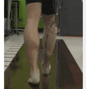
Gait Characteristics When Walking on Different Slippery Walkways
Mariah W. Whitmore, Levi J. Hargrove, Eric J. Perreault, Northwestern University, Rehabilitation Institute of Chicago, USA
Volume 63, Issue 1, Page: 228-239
Slippery surfaces are a dangerous, common cause of falling. This is especially true for individuals with a lower-limb amputation because current prostheses are unable to adapt to the slippery surfaces as intact legs do. We investigated how able-bodied subjects walked without slipping across different slippery walkways to better understand how a prosthesis should be controlled under these conditions. By characterizing changes in lower-limb muscle activity and kinematics of the able-bodied population we can gain an initial estimate of how a prosthetic limb should behave on slippery surfaces to minimize the users risk of slipping.