 Cardiac electrogram (EGM) signals and electrophysiologic (EP) characteristics derived from them such as amplitude and timing are central to the diagnosis and therapeutic management of arrhythmias. Bipolar EGMs are often used but possess polarity and shape dependence on catheter orientation contributing to uncertainty. We describe a novel method to map cardiac activation that resolves signals into more meaningful directions and is insensitive to electrode directional effects.
Multielectrode catheters that span 2- and 3-dimensional space are used to derive local electric field (E-field) signals. A traveling wave model of local EGM propagation motivates a new “omnipolar” reference frame in which to understand EGM E-field signals. This reference frame provides bipolar component EGMs aligned with anatomic and physiologic directions. We validate the basis of this technology and determine its accuracy using a saline tank in which we simulate physiologic propagation. Further, we verified omnipole signals from healthy tissue are nearly free of catheter orientation effects, are constrained by biophysics to consistent morphologies, and thus provide uniquely consistent amplitudes and timings.
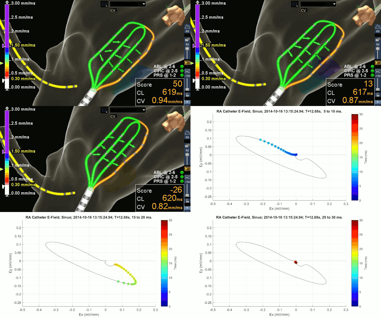
Using a 3D EP mapping system, traveling wave approximation, and Omnipolar Mapping Technology (OT) E-field loops, we demonstrate a new and nearly instantaneous means to determine local amplitude, activation time, conduction velocity and activation direction. OT’s catheters, approach to signal processing, and real-time visualization allow for a rapid and detailed characterization of myocardial activation that is insensitive to catheter orientation.

