
Thromboembolism in blood vessels can lead to stroke or heart attack and even sudden death unless brought under control. Despite the promising treatment results obtained via sonothrombolysis mediated with ultrasound contrast agents, the treatment efficacy for clinical application may not be optimized due to the lack of imaging guidance and clot characterization during the thrombolysis procedure. Therefore, this paper demonstrated a miniaturized transducer integrated with an optical fiber for photoacoustic-imaging (PA)-guided intravascular sonothrombolysis.
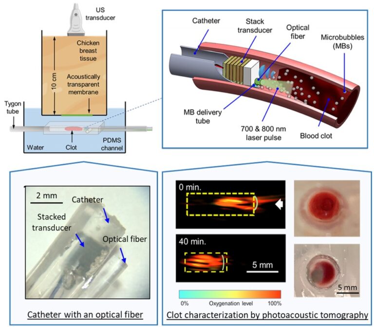
In specific, a miniaturized transducer composed of an 8-layer PZT-5A stack was developed with an aperture size of 1.4 ×1.4 mm2. Then, the transducer was assembled into a customized two-lumen 10-Fr catheter and integrated with an optical fiber with core diameter of 1.5 mm. After characterizing the performance of the transducer, the device was applied for the in-vitro microbubble-mediated sonothrombolysis tests. The treatment process was monitored with internal-illumination photoacoustic tomography (II-PAT), a hybrid imaging modality that combines the rich contrast of optical absorption and the deep penetration capability of ultrasound detection. Passive cavitation detection (PCD) was also conducted to examine the cavitation activities during sonothrombolysis.
The test results showed an improved lysis rate with microbubble-mediated sonothrombolysis accompanying with an enhancement of passive cavitation signals. With intravascular light delivery, II-PAT overcomes the penetration depth limited by strong optical attenuation of tissue. Clot position, shape, stiffness, and oxygenation level can be estimated by II-PAT at clinically relevant chicken tissue depth of about ten centimeters. For the first time, our findings have demonstrated the feasibility of the proposed PAT-guided intravascular sonothrombolysis with real-time feedback during the treatment process.