
Proper gastrointestinal motility is a requirement of a healthy digestive system, enabling the breakdown and transport of ingested nutrients. Bioelectrical ‘slow waves’ regulate these contractions, alongside other co-factors. We aimed to confirm whether the pyloric sphincter demarcates slow waves in the intact stomach and duodenum.
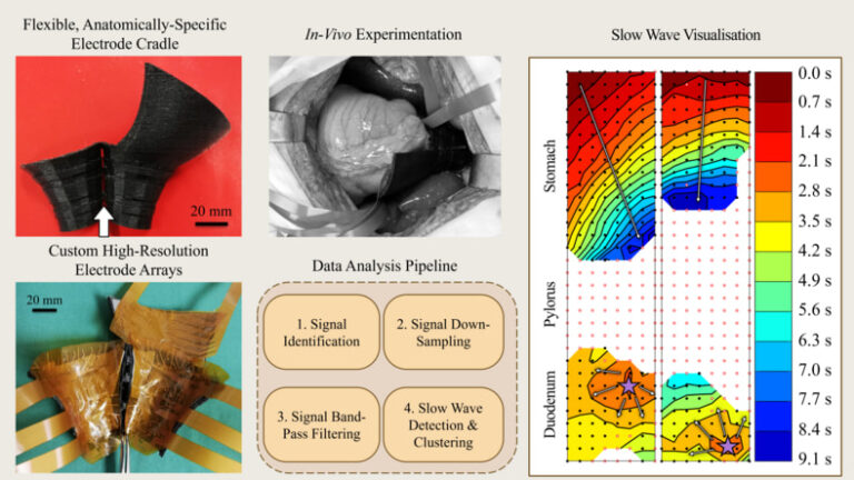
We developed and validated novel anatomically-specific electrode cradles and analysis techniques which enable in vivo high-resolution slow wave mapping of bioelectrical slow waves across the gastroduodenal junction. Cradles housed custom-fit, flexible electrode arrays during acute porcine experiments (N=9; 44.92 kg ± 8.49 kg) and maintained electrode contact with the gastroduodenal serosa. Simultaneous gastric and duodenal slow waves were filtered independently after determining suitable organ-specific filters. Validated algorithms calculated slow wave propagation patterns and quantitative descriptions.
Butterworth filters, with cut off frequencies (0.0167 – 2) Hz and (0.167 – 3.33) Hz, were determined to be the optimal filters for gastric and intestinal slow wave signals, respectively. There was a quiescent zone of (46.54 ± 5.73) mm wide in the pylorus, which separated gastric slow waves (2.76 ± 0.37) cpm and duodenal slow waves (18.13 ± 0.56) cpm. The duodenal slow waves originated from a pacemaker region (7.24 ± 4.70) mm distal to the quiescent zone. There was no temporal relationship between antral and duodenal slow waves across the pyloric region.
Novel engineering methods enable measurement of in vivo electrical activity across the gastroduodenal junction and provide qualitative and quantitative definitions of slow wave activity. The pylorus is a clinical target for gastrointestinal motility disorders and further investigations may reveal novel biomarkers. These biomarkers may enable diagnostic and treatment practices based upon organic cause, rather than relying on symptomatic profiles.