
Neovascularization of injured tendons prolongs the proliferative phase of healing, but prolonged neovascularization may cause improper healing and pain. Currently, ultrasound Doppler imaging is used for measuring the neovascularization of injured tendons (e.g., Achilles tendon). However, the resolution of state-of-the-art clinical ultrasound machines is insufficient for visualizing the neovascularization in finger tendons. In this study, a high-resolution micro-Doppler imaging (HFμDI) based on 40-MHz ultrafast ultrasound imaging was proposed for visualizing the neovascularization in injured finger tendons during multiple rehabilitation phases.
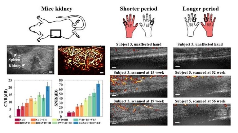
The vessel visibility was enhanced through a block-wise singular value decomposition filter and several curvilinear structure enhancement strategies, including the bowler-hat transform and Hessian-based vessel enhancement filtering. HFμDI was verified through small animal kidney and spleen imaging because the related vessel structure patterns of mice are well studied. Five patients with finger tendon injuries underwent HFμDI examination at various rehabilitation phases after surgery (weeks 11–56), and finger function evaluations were performed for comparisons.
The results of small animal experiments revealed that the proposed HFμDI provides excellent microvasculature imaging performance; The contrast-to-noise ratio was approximately 15 dB higher than the conventional methods, and the minimum detectable vessel size for mouse kidney was 35 μm without the use of contrast agent. In the human study, neovascularization was clearly observed in injured finger tendons during the early phase of healing (weeks 11–21), but it regressed from week 52 to 56. Finger rehabilitation appears to help reduce neovascularization; neovascular density decreased by approximately 1.8%–8.0% in participants after 4 weeks of rehabilitation. The experimental results verified the performance of HFμDI for microvasculature imaging and its potential for injured finger tendon evaluations.