
Estimating the neovascularity of human finger tendon through high-frequency ultrasound micro-Doppler imaging
https://www.embs.org/tbme/wp-content/uploads/sites/19/2022/07/TBME-02051-2021-Website-Image.jpg
780
435
IEEE Transactions on Biomedical Engineering (TBME)
//www.embs.org/tbme/wp-content/uploads/sites/19/2022/06/ieee-tbme-logo2x.png
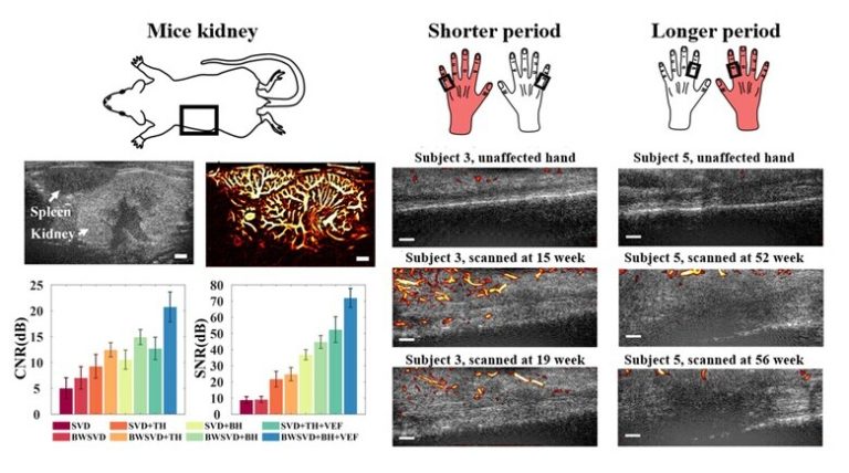
High-frequency micro-Doppler imaging (HFμDI) based on 40-MHz ultrafast ultrasound imaging was proposed for visualizing the neovascularization in injured finger tendons through block-wise singular value decomposition filtering and curvilinear structure enhancement. Small animal imaging experiments revealed that HFμDI provides excellent performance and the minimum vessel size was 35 μm without contrast agents. Neovascularization was clearly observed in injured finger tendons during the early phase of healing (weeks 11–21) and regressed from week 52-56. Neovascular density decreased by approximately 1.8%–8.0% after 4 weeks of rehabilitation. The experimental results indicate the potential of HFμDI for injured finger tendon evaluations.
read more