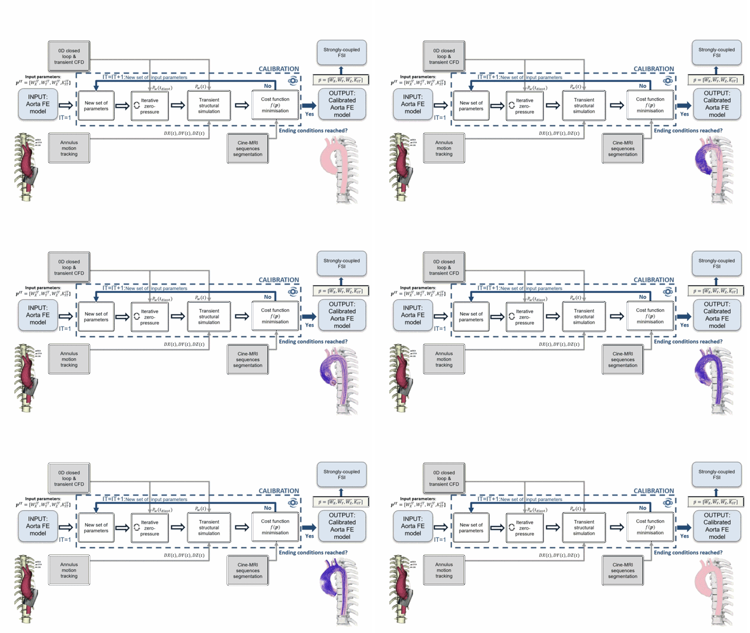
In this work, we proposed a procedure for calibrating 4 parameters governing the mechanical boundary conditions of a thoracic aorta model derived from one patient with ascending aortic aneurysm. The boundary conditions reproduced the visco-elastic structural support provided by the soft tissue and the spine and allowed for the inclusion of the heart motion effect.
The thoracic aorta was first segmented from magnetic resonance imaging (MRI) angiography and the annulus motion derived by 2D cine-MRI motion tracking. A rigid-wall fluid-dynamic simulation was performed to extract the time-varying wall pressure field. We built a Finite Element model considering patient-specific material properties and imposing the derived pressure field and the motion at the annulus boundary. The calibration, which involved the zero-pressure state computation to include the pre-stress, was based on purely structural simulations. After obtaining the vessel boundaries by segmenting the cine-MRI sequences, an iterative procedure was performed to minimize the distance between them and the corresponding boundaries derived from the deformed structural model. A strongly-coupled fluid-structure interaction analysis through a penalty-coupling method was finally performed with the tuned parameters and compared to the purely structural simulations.
The calibration with structural simulations allowed to reduce maximum and mean distances between image-based and simulation-derived boundaries from 8.64 mm to 6.37 mm and from 2.24 mm to 1.83 mm, respectively. The maximum root mean square error between the deformed structural and fluid-structure interaction surface meshes was 0.19 mm. This procedure could prove crucial for increasing the fidelity in replicating the real aortic root kinematics, potentially resulting in a more accurate assessment of properties such as stress and strain at the wall.

