
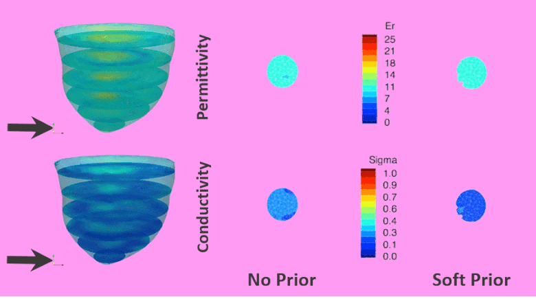
In this work, we present methods and results for fusion of magnetic resonance imaging(MRI) breast images with microwave tomography through a soft prior regularization technique. This method incorporates accurate boundary location of different regions of interest from MRI as spatial information into the regularization process of the image reconstruction algorithm. Methods: Numerical experiments were completed on a set of 3D breast geometries derived from MR breast data with different parenchymal densities, as well as a simulated tumor to evaluate performance over a range of breast shapes, sizes and property distributions. Results: When the soft prior regularization technique was applied, both permittivity and conductivity relative root mean square error (RRMSE) values decreased by more than 87% across all breast densities, except in two cases where the error decrease was only 55% and 78%. In addition, the incorporation of structural prior increased the recovered contrast between tumor and fibro-glandular tissue by 59% in permittivity and 192% in conductivity. Conclusion: This study confirmed that the soft prior regularization algorithm is robust in 3D microwave image reconstruction and can function successfully across a range of complex geometries and tissue property distributions. Significance: This study demonstrates that our microwave tomography is capable of recovering accurate tissue property distributions when spatial information from MRI is incorporated through soft prior regularization. Improvement is substantial and minimizes ambiguity at tissue interfaces. Gains were realized without the aid of priors on the property values, themselves.