
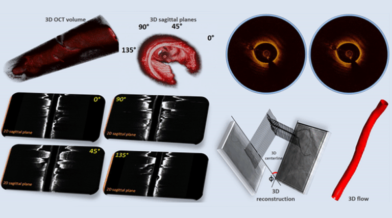
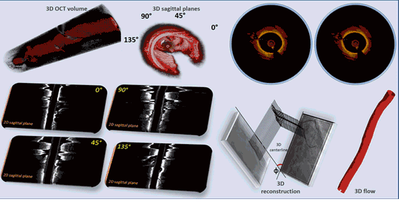
Real time segmentation and three-dimensional (3D) reconstruction of the coronary arteries is challenging and limited by the increased computational time of the developed computer-aided methods. We present a novel and time-efficient method for intracoronary lumen detection which produces 3D coronary arteries using Optical Coherence Tomographic (OCT) images. OCT images are acquired for multiple patients and longitudinal cross-section (LOCS) images are reconstructed using different acquisition angles. The lumen contours for each LOCS image are extracted and translated to 2D cross-sectional images. Using two angiographic projections the centerline of the coronary vessel is reconstructed in 3D and the detected 2D contours are transformed to 3D and placed perpendicular to the centerline, forming the vessel surface. To validate the proposed method, 613 manual annotations from medical experts were used as gold standard. The 2D detected contours were compared to the annotated contours and the 3D reconstructed models produced using the detected contours were compared to the models produced by the annotated contours. WSS, as dominant hemodynamics factor, was calculated using computational fluid dynamics and 844 consecutive 2-mm segments of the 3D models were extracted and compared to each other. There was an excellent agreement between the models produced by our method and by the experts’ annotations. The proposed computer-aided method is fast, accurate and likely to assist in a number of research and clinical applications.