
Biological tissues such as the myocardium or the artery walls are subject to fast and complex phenomena which are related to their physical properties (geometry, composition, stiffness, etc.) as well as their function. Imaging these characteristics is essential from a medical point of view to pose a diagnosis or to understand the development of some diseases. However, even if new motion estimation techniques and algorithms have been developed and improved in the past years, the visualization method is still a challenge to render and represent tissues and their motion in an accurate and intelligible manner.
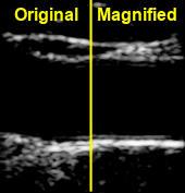
In the meantime, video magnification, which is a computational method increasing the subtle temporal motion in videos, has been developed for vision applications; small changes are amplified to be easily seen with a naked eye. Herein, video magnification has been applied to medical ultrasound images.
The primary purpose of the study was to develop a visualization technique for tissue motion, using video magnification, to get rid of color maps and vectors. At this end, carotid acquisitions were performed using an ultrasound scanner and motion was extracted using a Doppler technique before video magnification. Thanks to video magnification, local displacement, and distortion (shear stress) as well as complex and fast phenomena (pulse wave) can be seen with the naked eye.
The method can be applied to any biomedical dynamic imaging modality as long as the temporal resolution and motion estimation technique used is suitable to the phenomenon of interest. Video magnification could be a new tool for physicians to highlight new pathology indicators or for long-term disease monitoring; it could also be used for rapid qualitative inspection or educational purposes.