
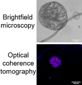
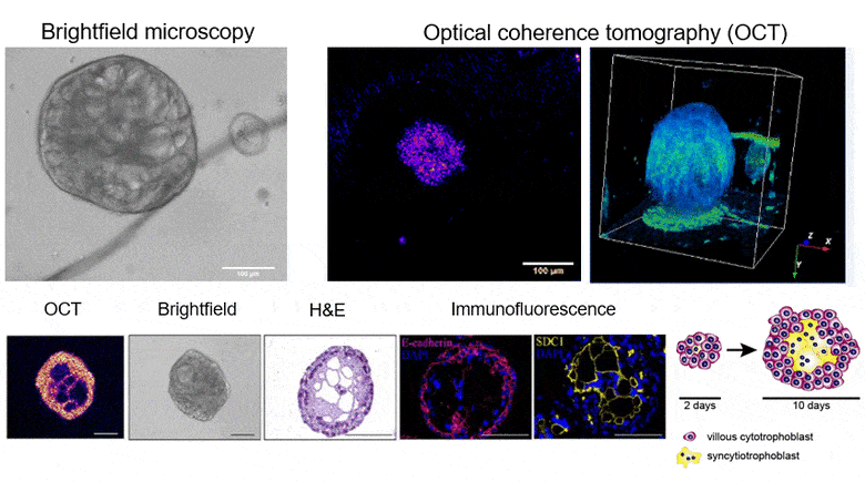
Correct placental development/function plays a crucial role in maternal and fetal health not only during pregnancy but also later in life. Recently, placenta-derived trophoblast organoids (TB-ORG) have been developed. Organoids are 3D multicellular structures which closely recapitulate certain formations and functions of individual organs. The establishment of TB-ORG was a milestone in trophoblast research allowing to investigate physiological and pathophysiological placental development, differentiation, and function in vitro. However, morphological investigations of TB-ORG architectures involve the fixation of TB-ORG and subsequent immunohistochemical analyses. These conventional methods require the termination of individual experiments and leads to morphological modifications of TB-ORG structures. Brightfield microscopy indeed allows to depict the overall organoid morphology but cannot resolve their inner structures that is composed of differentiated, multinucleated, hormone-producing syncytiotrophoblasts (STBs). To address these shortcomings, we introduced optical coherence tomography (OCT) for imaging TB-ORGs during cultivation. OCT is based on low-coherence interferometry and intrinsic scattering contrast from the sample. This article covers the results obtained with a novel polarization aligned ultra-high-resolution spectral domain 3D OCT featuring a central wavelength of 846 nm, 3 dB bandwidth of 133.3 nm, and uniform resolution of 2.48 μm in organoids. We herein demonstrate for the first time, that OCT can image the inner structures and 3D orientation of TB-ORG without sample labeling and/or destruction. Furthermore, we were able to detect “bridges” and cavities within the organoids which were signs for STB formation. These findings were verified by detecting specific STB markers using quantitative polymerase chain reaction, Western blot analyses, and immunofluorescence. In summary, OCT enables a more detailed depiction of TB-ORG structures compared to brightfield microscopy. Furthermore, OCT allows to determine the STB differentiation state of individual TB-ORG without sample destruction and/or labeling. Hence, OCT may become an important imaging tool for TB-ORG studies.