
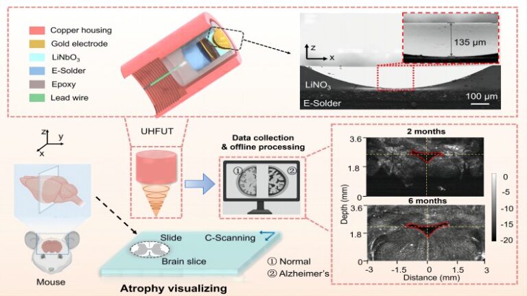
Characterizing mouse brain anatomy is crucial for investigating brain injuries and degenerative diseases in mouse models. Achieving optimal spatial resolution and imaging depth is essential for enhancing in vivo imaging of embryonic and mouse brains. To address these challenges, we present an efficient approach utilizing ultra-high frequency self-focusing ultrasonic sensors with ultra-broad bandwidth to visualize mouse brain atrophy.
The sensor, fabricated through micro-nano processing technology, features a half-concave element. Numerical models, based on the differential method, provide geometry and theoretically predicted acoustic parameters for these elements. Demonstrating the sensor’s efficacy, we assessed structural changes in mouse brain slices at different ages, revealing excellent imaging performance that captures atrophy in aged mouse brains. With a lateral resolution of 24 μm and a bandwidth of 115% at -6 dB, we imaged 1 mm thick brain slices from 2 and 6-month-old mice using a C-mode acoustic microscopy system. A comparison highlighted significant width differences in the 4th ventricle (4V) between older and younger mice. Combining brain atrophy visualization with neuroscience holds potential for diagnosing and treating Alzheimer’s disease (AD). Additionally, acoustic properties of brain slices were quantitatively measured, though the sub-millimeter thickness and averaged calculations warrant future exploration with higher frequency sensors and thinner slices for more accurate characterization. This method holds significance for high-resolution imaging of biological tissues with ultra-high frequency self-focusing ultrasonic sensors.
In conclusion, these promising results showcase the potential application of ultra-high frequency self-focusing ultrasonic sensors for high-resolution in vitro imaging of biological tissues, particularly in studying mouse brain anatomy and addressing neurodegenerative diseases like AD.