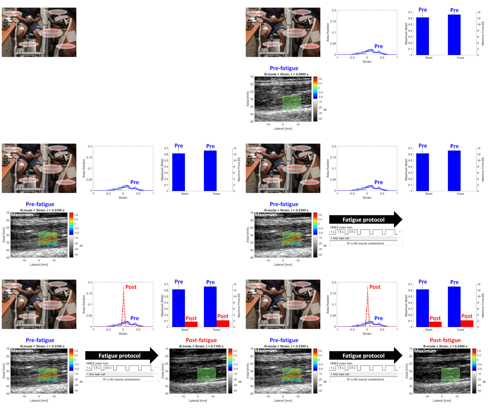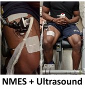
Neuromuscular electrical stimulation (NMES) has been demonstrated to activate paralyzed or paretic muscles that in turn help restore limb functions (e.g., standing, walking, upper limb functions such as grasp and reaching) of persons with stroke, hemiplegia, paraplegia and other neurological disorders. However, it is commonly noted that during NMES operation time, a rapid onset of muscle fatigue quickly deteriorates the force generation capability of the muscle and limits the effectiveness of NMES applications. This study investigates a novel ultrasound imaging-based non-invasive methodology to quantitatively assess changes in muscle contractility due to the fatigue induced by the NMES. Isometric knee extension experiments on human participants were conducted before and after a designed NMES fatigue protocol. Under a synchronized data acquisition configuration, the force produced by the muscle contraction was registered by a load cell at 1 kHz while the ultrasound images were acquired by a 2 kHz ultra-fast plane wave imaging mode. By comparing the experiment data before and after the fatigue protocol, we first showed a significant reduction of the force that is used to justify the applied fatigue protocol. A two-dimensional time-variant strain field along the time course of the muscle contraction was then derived by processing the ultrasound images using a contraction-rate adaptive speckle tracking algorithm. Analysis of the derived strain images showed that, between the pre-fatigue and post-fatigue stages, there were a significant reduction in the strain peaks, a change in the strain peak distribution, and a significant decrease in an area occupied by the large positive strain. The results indicate that ultrasound imaging is promising to assess changes in muscle contractility due to the NMES-induced muscle fatigue. The capability of a direct fatigue assessment is a critical precursor to design the optimal NMES dosage in control of a neuroprosthesis that can alleviate the NMES-induced fatigue effect.

