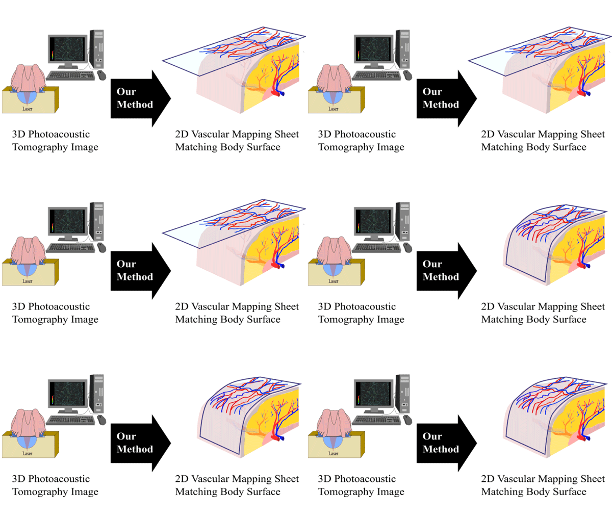
Flap surgery is a medical procedure that involves the transfer of a living piece of tissue, with an intact blood supply, from one part of the body to another (e.g. areas that have lost skin, fat, muscle or skeletal support). Equally critical to successful skin flap transfer is the understanding of the skin’s blood supply architecture and the identification of the dominant perforator vessel in the flap. However, the exact location of the perforators varies significantly among patients.
Photoacoustic tomography (PAT), that permits rapid three-dimensional (3D) mapping of blood vessel architecture, is a promising imaging system for preoperative planning of perforator flaps. In this study, we introduce a new protocol to create two-dimensional (2D) vascular mapping sheets that can support the preoperative planning. The core of this protocol is PreFlap, a new algorithm that unwraps 3D blood vessel points from PAT images into 2D vascular mapping sheets taking into account the shape of the body surface. These sheets can be overlaid on the body and help the surgeon to identify vessels and perforators even during the surgery.
We conducted phantom and clinical studies reporting the complete image processing from the 3D PAT image data acquisition to the 2D mapping sheet creation. Our results not only validated the PreFlap algorithm and demonstrated the reduction of measurement errors associated with the vessels localization, but also revealed that PreFlap can improve preoperative flap evaluation saving surgeons’ time and improving surgical outcomes.

