Kamil Uğurbil, University of Minnesota, Volume 61, Issue 5, page: 1364-1379


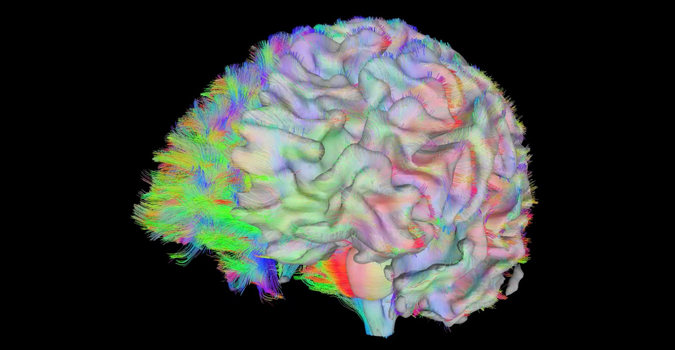
The last two decades have witnessed revolutionary changes in our ability to study biological processes in the human body, in particular the human brain. Magnetic resonance Imaging (MRI) and spectroscopy at high magnetic fields (3 Tesla and above) has played a critical role in this development. The introduction of MRI methods to image human brain function (fMRI) in 1992 and the realization of the strong magnetic field dependence of these functional signals have ultimately led to the introduction of 7 Tesla for human studies at about the turn of the century. Work conducted at this ultrahigh magnetic field demonstrated the existence of significant advantages in sensitivity and biological information content, as well as the presence of numerous challenges. With the development of engineering and methodological solutions to these challenges, ultrahigh fields have provided unprecedented gains in imaging human brain activity, spanning the scales of brain organization ranging from elementary computational units to whole brain networks, and have started to make inroads into the investigation of other organ systems. As extensive as they are, these gains still constitute a prelude to what is to come given the increasingly larger effort committed to ultrahigh field research, the anticipated introduction of 10.5 and 11.7 Tesla systems for human imaging, and the incessant developments of better instrumentation and imaging techniques.

