
Ultrasound technology has increasingly been applied to modulate neural activity and can elicit both excitatory and suppressive effects when applied to neural brain tissue, however, few studies have studied its effect in spinal cord tissue. In this study, we investigated the ability of low-intensity ultrasound (LIUS) applied to the spinal cord to modulate the transmission of motor signals in a rodent model.
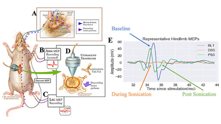
We utilized ten male adult Sprague-Dawley rats (250-300 g, 15 weeks old). Cranial electrodes were placed for stimulating motor evoked potentials (MEPs), while upper and lower extremity electrodes were placed for recording. A thoracic laminectomy was performed to expose the spinal cord at the T11 and T12 vertebral levels. A LIUS transducer placed directly above the exposed spinal cord sonicated the cord for either 5 or 10 minutes while MEPs were acquired each minute. Following the sonication period, the ultrasound was turned off, and post-sonication MEPs were acquired for an additional 5 minutes. We found that hindlimb MEP amplitude significantly decreased during sonication in both the 5- (p<0.001) and 10-min (p=0.004) cohorts followed by a gradual return to baseline, while forelimb MEP amplitude did not demonstrate any statistically significant changes. Histology and qPCR demonstrated that sonication did not cause damage to the tissue. Ex vivo temperature analysis indicated minimal change in tissue temperature during sonication.
Our study showed that LIUS applied to the spinal cord suppresses MEP signals caudal to the site of sonication, with recovery of MEPs to baseline after sonication. These findings may have translation potential in treating movement disorders driven by excessive excitation of spinal neurons.