
Laparoscopic surgery, a minimally invasive technique, offers advantages like reduced scarring and quicker recovery times but presents challenges in visualizing critical structures like nerves and blood vessels, leading to potential complications. This study focuses on developing a miniaturized laparoscopic photoacoustic (PA) imaging system to enhance neurovascular detection during surgery by leveraging their unique optical absorption properties.
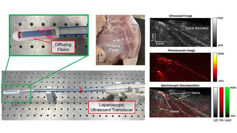
Our approach involved modifying a commercially available ultrasound laparoscopic probe by integrating custom-fabricated side-illumination diffusing fibers to illuminate the probe’s field of view. We successfully measured and modeled the light distribution emitted from these diffusing fibers. Optical simulations were employed to replicate the light distribution and assess how fiber alignment influences the penetration depth of the PA signal, ultimately determining optimal alignment angles for maximizing PA signal quality at various depths.
We conducted phantom and ex vivo studies to validate the system’s imaging capabilities. In the phantom study, our system was tested in imaging targets submerged within an optically scattered medium, consistently achieving an average lateral full-width at half-maximum of 0.43±0.09 mm and a signal-to-noise ratio of 31.2±1.84 dB for 0.2 mm diameter wire at depths ranging from 6.5 to 26.5 mm. In the ex vivo study, our system was tested with rat thigh. The tissue boundary was accurately captured in PA imaging and aligned with the location of the US-scanned sample surface. The PA image captured several subsurface features, such as vessels and nerves. The vessel can be clearly visualized at a depth of 13.27 mm from the transducer.
These results underscore the potential of a side-illumination diffusing fiber PA imaging system for guidance in laparoscopic surgery, with the clinical translation of this technology holding promise for preserving critical vascular structures and nerves and thereby reducing post-operative complications.