Volume 62, Issue 5, Page: 1416-1424

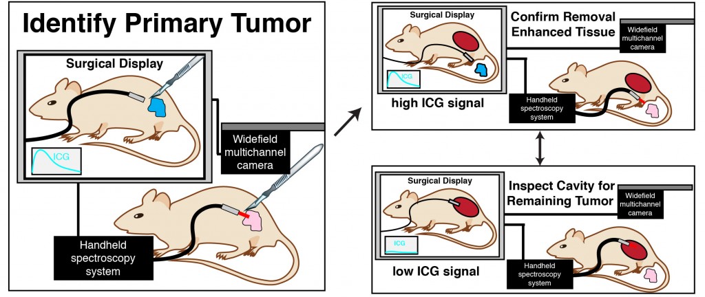
Surgery is the most widely used treatment for solid tumors worldwide. However, recurrence of the tumor following surgery continues to prevent many patients from achieving complete remission. There are several factors that contribute to tumor recurrence; the goal of this work was to develop and evaluate an image-guided surgery system that could detect the presence of residual tumor remaining at or near the surgical site during surgery. We developed a near infrared system that employs a handheld excitation source and spectrometer in combination with a widefield video imaging system. After injection of a near infrared fluorescent imaging agent, in this case, indocyanine green (ICG), the imaging system was evaluated for its optical performance and ability to detect the presence of indocyanine green in tumors. We tested the ability of the system to detect ICG in a mouse model of triple negative breast cancer as well as in spontaneous tumors arising in canines, in this case fibrosarcoma. In both examples, an intravenous bolus of ICG resulted in significant accumulation in tumor or neoplastic tissue relative to surrounding normal tissue. The significant accumulation of ICG in tumor could be readily detected by the image-guided surgery system and low accumulation by surrounding tissue resulting in strong contrast under video guidance. This work highlights the potential of local, directed excitation with point spectroscopy in combination with widefield imaging. Moreover, the ability to readily detect ICG in canines with spontaneous tumors in a clinical setting exemplifies the potential for further clinical translation.

