
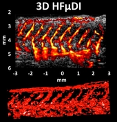

Zebrafish has been recently considered an ideal vertebrate for studying developmental biology, genetics, particularly for modeling tumorigenesis, angiogenesis, and regeneration in vivo. However, when a zebrafish matures completely, particularly if the specimen is from a wild-type line, stripes are evident along the length of the body; such stripes reduce transparency. Optical imaging may have limitations for adult zebrafish imaging because of its low penetration ability. Acoustic wave penetration outperforms optical methods, high-frequency (>30 MHz) ultrasound (HFUS) was consequently an alternative imaging modality for adult zebrafish imaging, particularly for echocardiography However, visualizing peripheral vessels in a zebrafish by using conventional HFUS is still difficult. In the present study, high-frequency micro-Doppler imaging (HFμDI) based on ultrafast ultrasound imaging was proposed for adult zebrafish dorsal vascular mapping in vivo. HFμDI uses a 40-MHz ultrasound transducer, which is an ultrafast ultrasound imaging technology with the highest frequency available currently. Blood flow signals were extracted using an eigen-based clutter filter with different settings. Experiments were performed on an 8-month-old wild-type AB-line adult zebrafish. Blood vessels, including intersegmental vessels, parachordal vessel, dorsal longitudinal anastomotic vessel, and dorsal aorta, from the dorsal side of the adult zebrafish were clearly observed in two-dimensional (2D) and three-dimensional (3D) HFμDI. The maximum image depth of HFμDI and the minimal diameter of vessel can be detected were 4 mm and 36 μm, respectively; they were determined without any use of microbubbles. The maximum flow velocity range was approximately 3–4 mm/s on the dorsal vessels of the adult zebrafish.