
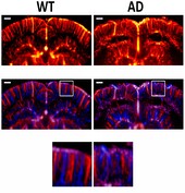
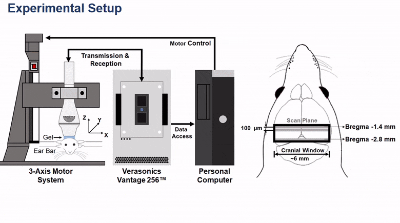
Objective: Cerebrovascular disorders are associated with Alzheimer’s disease (AD). Preclinical animal study is necessary for understanding AD pathogenesis and determining its optimal diagnosis and treatment strategies. Conventionally, the cerebral vasculature’s structure is analyzed through histological staining. However, functional analysis of the cerebral vasculature requires an in vivo approach to visualize the blood flow in small animal brains. Methods: This paper proposes high-frequency micro-Doppler imaging (HFμDI) technology for mapping mouse cerebral vasculature. Triple-transgenic mice, expressing three familial AD mutant genes (PS1M146V, tauP301L, and APPSwe), were obtained from Jackson Laboratory (Bar Harbor, ME, USA) and used as model AD mice. Nongenetically modified C57BL/6 mice were used as WT mice. The cerebral circulation in the cortex and hippocampus was visualized through HFμDI, and the vessel density and anatomy of the regions were calculated using the images. Results: Using a 40-MHz transducer enabled in vivo visualization of the mouse brain up to 3 mm in depth; furthermore, a minimal vessel diameter of 48 μm could be determined without using microbubbles. The optimal HFμDI setting was confirmed by calculating a vessel image’s contrast. The excited pulse is a three-cycle bipolar signal, and the tilted angle is 7°. A frame rate of 1 kHz can be achieved using this setting. Animal experiments determined that the cortical and hippocampal vessel density in young (4-month-old) wild-type mice was similar to that in middle-aged (11-month-old) wild-type mice. However, compared with the vessel density in middle-aged wild-type mice, that in middle-aged mice with AD was significantly lower, particularly in the hippocampus. Conclusion: In vivo observation of cerebral vasculature demonstrated the effectiveness of HFμDI for the preclinical study of AD and as a potential tool for AD diagnosis.