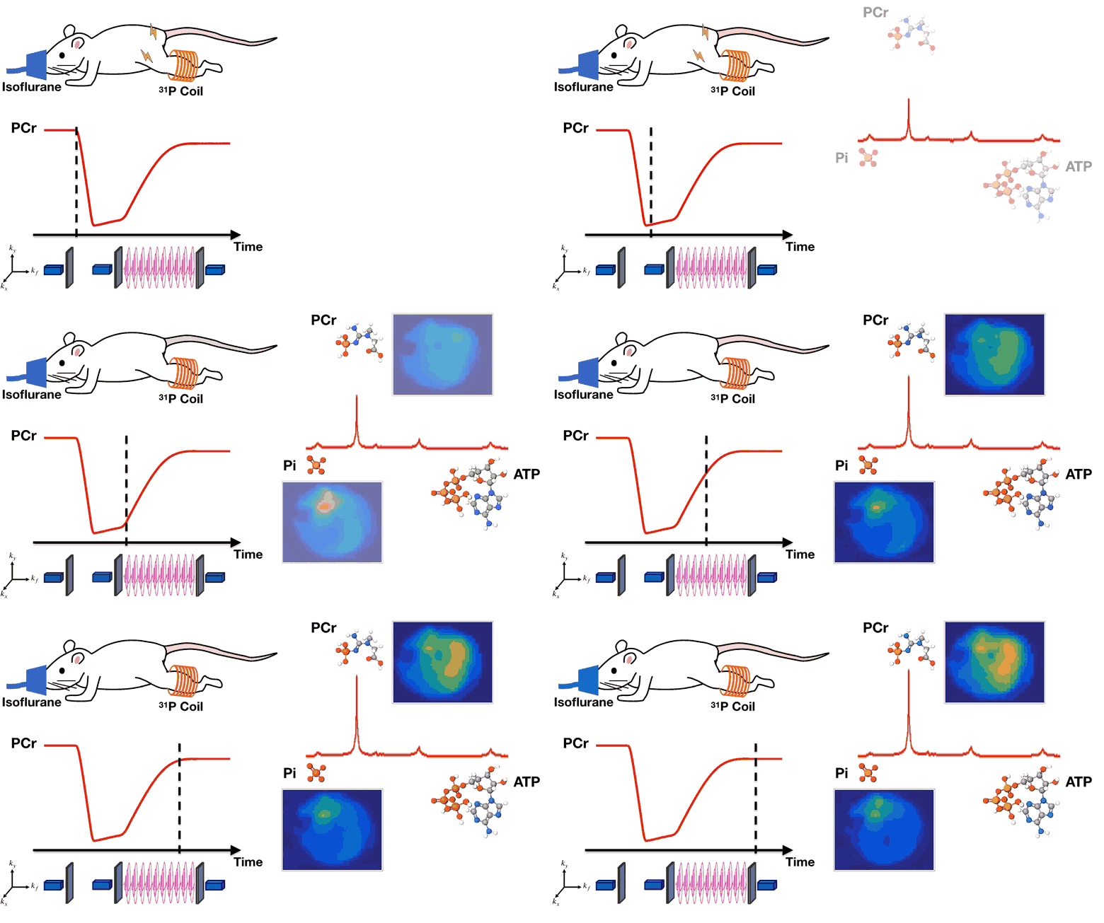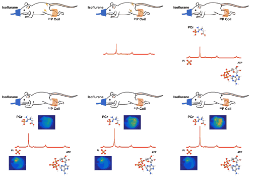
Abnormal mitochondrial metabolism is a hallmark of many prevalent diseases such as diabetes and cardiovascular disease; however, current understanding of mitochondrial function is mostly gained from studies on isolated mitochondria under nonphysiological conditions. To address this issue, we have developed a novel high-resolution 31P magnetic resonance spectroscopic imaging method which enables noninvasive dynamic mapping of several important phosphate metabolites such as phosphocreatine (PCr), ATP, and inorganic phosphate (Pi). The method synergistically integrates physics-based models of spectral structures, biochemical modeling of molecular dynamics, and subspace learning into a low-rank tensor imaging framework to capture the dynamic changes in phosphate metabolites during a metabolic perturbation protocol. Fast data acquisition is achieved by using rapid spiral trajectories and sparse sampling of the 5D spatial-spectral-temporal -space. Image reconstruction is accomplished using an efficient tensor recovery algorithm.
In vivo and in vitro experiments were performed to demonstrate the method’s ability to produce high-resolution dynamic metabolic mapping in rat hindlimb at spatial, spectral, and temporal resolutions of 4 4 2 mm3, 0.1 ppm, and 1.28 s, respectively. Multiple rounds of in vivo experiments were performed to demonstrate reproducibility, and in vitro experiments were used to validate the accuracy of the estimated metabolite maps. The high spatiotemporal resolution of the metabolite maps allowed for mapping of PCr resynthesis rates, a well-established index of mitochondrial oxidative capacity. Observed resynthesis rates were in agreement with literature values and regional analyses demonstrated differences between different muscle types.
These results demonstrate the utility of the proposed method for assessing mitochondrial function in vivo. This noninvasive method is highly translatable to clinical investigations and has the potential to open new avenues of research for studying disease progression and evaluating therapeutic intervention.

