
Ultrasound transient elastography (TE) technologies for liver stiffness measurement (LSM) are used clinically to screen for liver fibrosis. They utilize vibration of small, flat pistons, which generate shear waves that lack directivity and diverge away from the LSM region. The most common cause for LSM failure in practice is insufficient shear wave signal at the needed depths, especially in obese patients that are most at risk of liver disease. We propose a method to increase shear wave amplitude by focusing the waves into a directional beam that converges towards the LSM region.
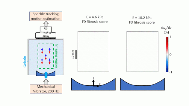
Here, we investigate the generation and propagation of focused shear wave beams (fSWBs) in liver tissue-mimicking gelatin. Directional fSWBs are generated by vibration at 200-400 Hz of a concave piston embedded near the surface of gelatin phantoms and measured with high-frame-rate ultrasound imaging. Five phantoms with a range of stiffnesses covering healthy and fibrotic liver are employed. Shear wave speeds assessed by fSWBs are compared with those by radiation-force-based methods (2D SWE). fSWB amplitudes are compared to predictions using a novel analytical model for shear wave generation and propagation.
We find that fSWB-derived shear wave speeds are in good agreement with 2D SWE. The amplitudes of fSWBs are localized to the LSM region and are significantly greater than unfocused shear waves. Overall agreement with theory is observed, with some discrepancies in the theoretical source condition. Challenges for translation include coupling piston vibration with the patient skin and increased shear wave attenuation in vivo compared to the phantoms employed here. Fibrosis is the most predictive measure of patient outcome in non-alcoholic fatty liver disease, and increased shear wave amplitude in the LSM region can reduce LSM failure rates by TE, thus reducing the need for invasive methods like biopsy or more expensive magnetic resonance elastography.