
Jin Keun Seo and Eung Je Woo, Yonsei University & Kyung Hee University,
Open Access, Volume 61, Issue 5, Page:1390-1399
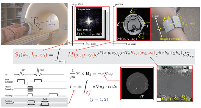
The tomographic imaging of tissue’s electrical properties (e.g., conductivity and permittivity) has been greatly improved by recent developments in magnetic resonance (MR) imaging techniques, which include MR electrical impedance tomography (MREIT) and electrical property tomography (EPT). When biological material is subjected to an external electric field, local changes in its electrical properties become sources of magnetic field perturbations, which are detectable by the MR signals. Controlling the external excitation and measuring the responses using an MRI scanner, we can formulate the imaging problem as an inverse problem in which unknown tissue properties are recovered from the acquired MR signals. This inverse problem is nonlinear; it involves the incorporation of Maxwell’s equations and Bloch equations during data acquisition. Each method for visualizing internal conductivity and permittivity distributions has its own methodological limitations, and is restricted to imaging only a part of the ensemble or mean tissue structures or states. Therefore, imaging methods can be improved by developing complementary methods that can employ the beneficial aspects of various existing techniques. This paper focuses on recent progress in MREIT and discusses its distinct features in comparison with other imaging methods. In MREIT, we inject low-frequency currents into an imaging object and measure the induced magnetic flux density data using an MRI scanner. Solving the nonlinear inverse problem in MREIT, we reconstruct cross-sectional images of the internal conductivity distribution. Potential applications of MREIT include tumor imaging and neuroimaging. It may also provide quantitative conductivity information to neuronal current source imaging and numerous therapeutic methods including tDCS and DBS.