
Three- (3-) and four-dimensional (4D) endorectal ultrasound are now available from most major ultrasound equipment manufacturers, but have been rarely deployed within prostate biopsy guidance systems despite the benefit of rapid volumetric imaging. The most common type of 3/4D transducers rotate the transducer array inside the probe casing using a small motor. However, in addition to the potential interference from certain metallic objects and electrical devices found in clinical environments, such motors are known to cause substantial distortion to tracking measurements made using some electromagnetic (EM) position sensors. Although EM tracking is becoming increasingly used in commercial systems for localising the transducer position with respect to the prostate, and for acquiring 3D volumes using a conventional two-dimensional (2D) transducer, supporting 3/4D transducers is problematic.
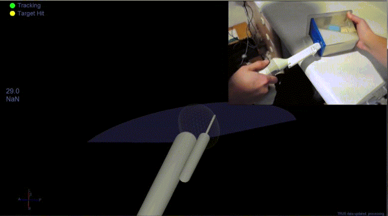
We describe the design, development and validation of a 3D-ultrasound-guided transrectal prostate biopsy system that employs high-accuracy optical tracking as an alternative to EM tracking to localise the ultrasound probe and prostate targets in 3D physical space. The system software incorporates an intuitive navigation method, based on realtime 3D graphical display of a virtual probe and biopsy needle trajectory, as well as a deformable MRI-ultrasound image registration algorithm that uses a combined statistical-biomechanical model of patient-specific prostate geometry and motion. A new clinical workflow specifically for the system is also described.
The system accuracy was validated by evaluating the targeted needle placement error after inserting a biopsy needle to sample planned targets in a phantom using standard 2D B-mode ultrasound guidance versus the proposed real-time 3D guidance. The overall mean (±SD) needle-segment-to-target and needle-to-target distance errors obtained using the new system were 3.6±4.0 and 3.2±2.4 mm, respectively. Furthermore, a significant increase in needle placement accuracy was observed when using the 3D guidance compared with visual targeting of invisible (virtual) lesions using a conventional ultrasound-guided biopsy technique.
Keywords: prostate cancer, image-guided interventions, image registration, tracking, transrectal biopsy