
Surgical staplers are commonly used in numerous open and laparoscopic cancer resection procedures including thoracic, bariatric, and colorectal surgery. In these procedures, it can be challenging to identify important tissues including lung nodules, artery branches, and lymph nodes. In particular, localization of small lung nodules during surgery may be thoracoscopically invisible and impalpable, requiring different localization methods. While current localization methods perform with high success rates, each approach has drawbacks and can fail in surgery. Thus, a tool that could provide intraoperative, real-time feedback on lung nodules or general tissue characterization could improve surgical outcomes. This study explores the feasibility of one such solution to this problem via coupling Electrical Impedance Tomography (EIT) to a standard-of-care laparoscopic surgical stapler, stapler+EIT. Electrical impedance is leveraged here as numerous ex vivo studies have confirmed a strong electrical property contrast between different tissue types, and notably between benign and malignant tissues.
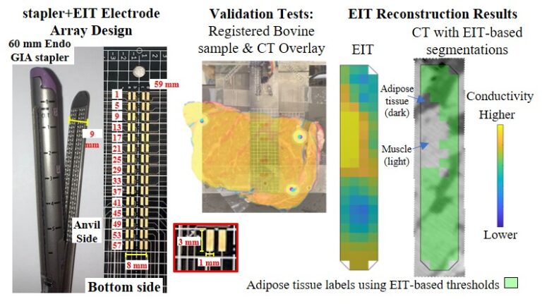
Two custom printed-circuit-board-based electrode arrays with 60 and 8 electrodes, respectively, matching the stapler geometry, served as the jaws of an electrode-integrated surrogate stapler+EIT device. The device was evaluated through a series of simulations and bench-top imaging experiments of agar-gel phantoms and bovine tissue samples to evaluate the technique and determine optimal imaging parameters.
Electrodes localized to only one jaw (the 60-electrode side) of the stapler outperformed a dual-jaw distribution of electrodes. Using this one-sided electrode array, reconstructions achieved an Area-Under-the-Curve (AUC) ≥0.94 for inclusions with conductivity contrasts of ≥30% for any size considered on measured agar-gel tests, and an AUC of 0.80 for bovine tissue samples. Overall, this study evaluated a stapler+EIT algorithm that has been tuned and evaluated on challenging phantom tests and demonstrated an ability to produce accurate reconstructions. This work is an important step in the development of a surgical stapler+EIT technique for margin assessment and/or tissue characterization.