Christian Bauer, Melissa A. Krueger, Wayne J. Lamm, Brian J. Smith, Robb W. Glenny, and Reinhard R. Beichel, University of Iowa – ECE
Volume 61, Issue 1, Page: 119-130

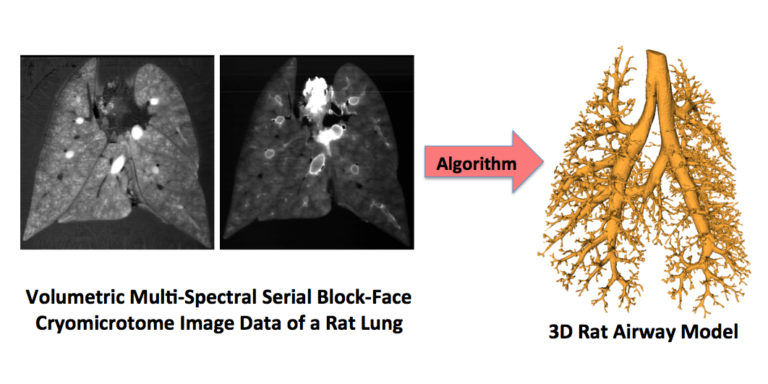
Serial block-face cryomicrotome imaging of rat lungs provides high-resolution information about lung anatomy as well as maps of regional ventilation and perfusion. The paper presents a novel highly-automated method for the segmentation of airways in cryomicrotome image data of rat lungs. First, a set of candidate airway centerline points is automatically identified. By utilizing a path extraction method, a centerline path between the root of the airway tree and each point in the set of candidate centerline points is obtained. Local disturbances are robustly handled by a path extraction approach, which avoids the shortcut problem of standard minimum cost path algorithms. The union of all centerline paths is utilized to generate an initial airway tree structure, and a pruning algorithm is applied to automatically remove erroneous subtrees or branches. Second, a surface segmentation method is used to obtain the airway lumen. The method was validated on five image volumes of Sprague–Dawley rats. The average of airway detection sensitivity was 87.4% with a 95% confidence interval (CI) of (84.9, 88.6)%, and a plot of sensitivity as a function of airway radius is provided in the paper. The combined estimate of airway detection specificity was 100% with a 95% CI of (99.4, 100)%. The average number and diameter of terminal airway branches was 1179 and 159 μm, respectively. Airway segmentation results include airways up to 31 generations. The presented approach enables quantitative studies of physiology and lung diseases in rats, requiring detailed geometric airway models.

