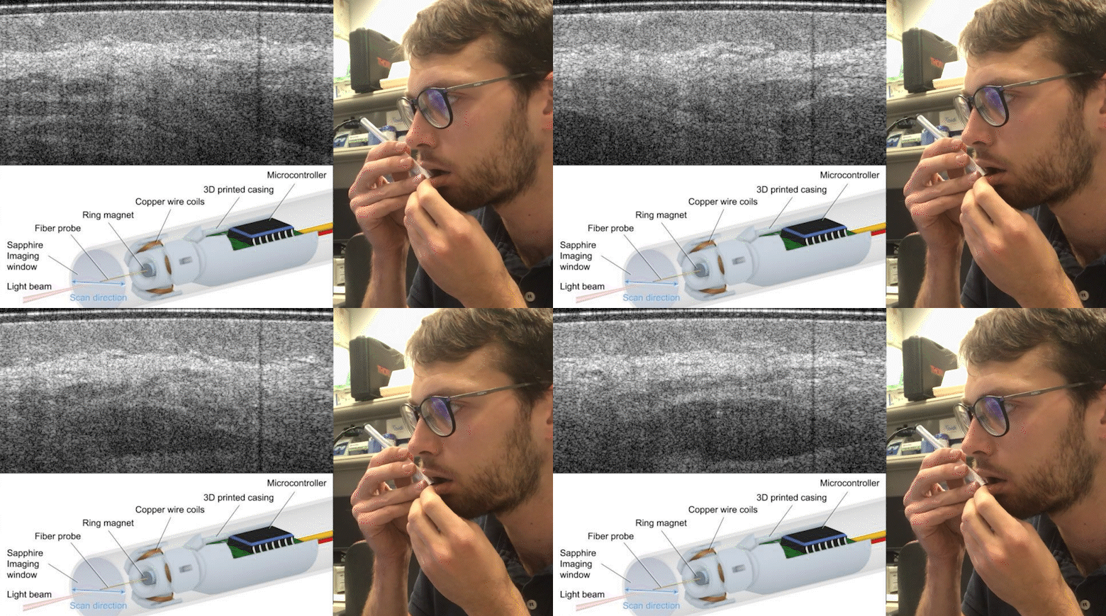
The soft tissue of the oral cavity is subject to a wide range of malignant and benign lesions that result in changes to the tissue microarchitecture. Optical coherence tomography (OCT) is non-invasive, subsurface imaging modality that is well-suited to visualizing pathological changes in tissue structure. However, OCT scanning probes tend to be large and bulky and unsuitable for use inside the mouth. In this study, we present the design of a small, light-weight handheld imaging probe that enables easy access to areas such as the mouth.
The probe uses an all-fiber optical design, in which a small lens (125 microns diameter) is fabricated on the distal end of an optical fiber to emit a weakly-focused, near-infrared light beam, and detect the light back-scattered from the tissue. The fiber is actuated using a magnetic scanning mechanism, in which a small rare-earth ring magnetic (1mm outer-diameter) is affixed around the fiber. This is mounted within a 3D printed casing and the fiber is scanned back-and-forth in the magnetic field generated by sinusoidally-charged copper wire coils to acquire a two dimensional OCT image at a rate of 50 images per second. The entire probe is encased within a small stainless-steel cylinder, approximately the size of a pen (10mm x 140mm).
We present results from six patients under-going examination for oral lichen planus, and one healthy control. Our results showed that OCT is able to visualize the loss of delineation between the epithelium and lamina propria in diseased tissue, and contrast these against the intact layered tissue structures in adjacent normal regions. We also demonstrate the ability of this small probe to acquire a freehand 3D scan by manually moving the probe across the tissue.
