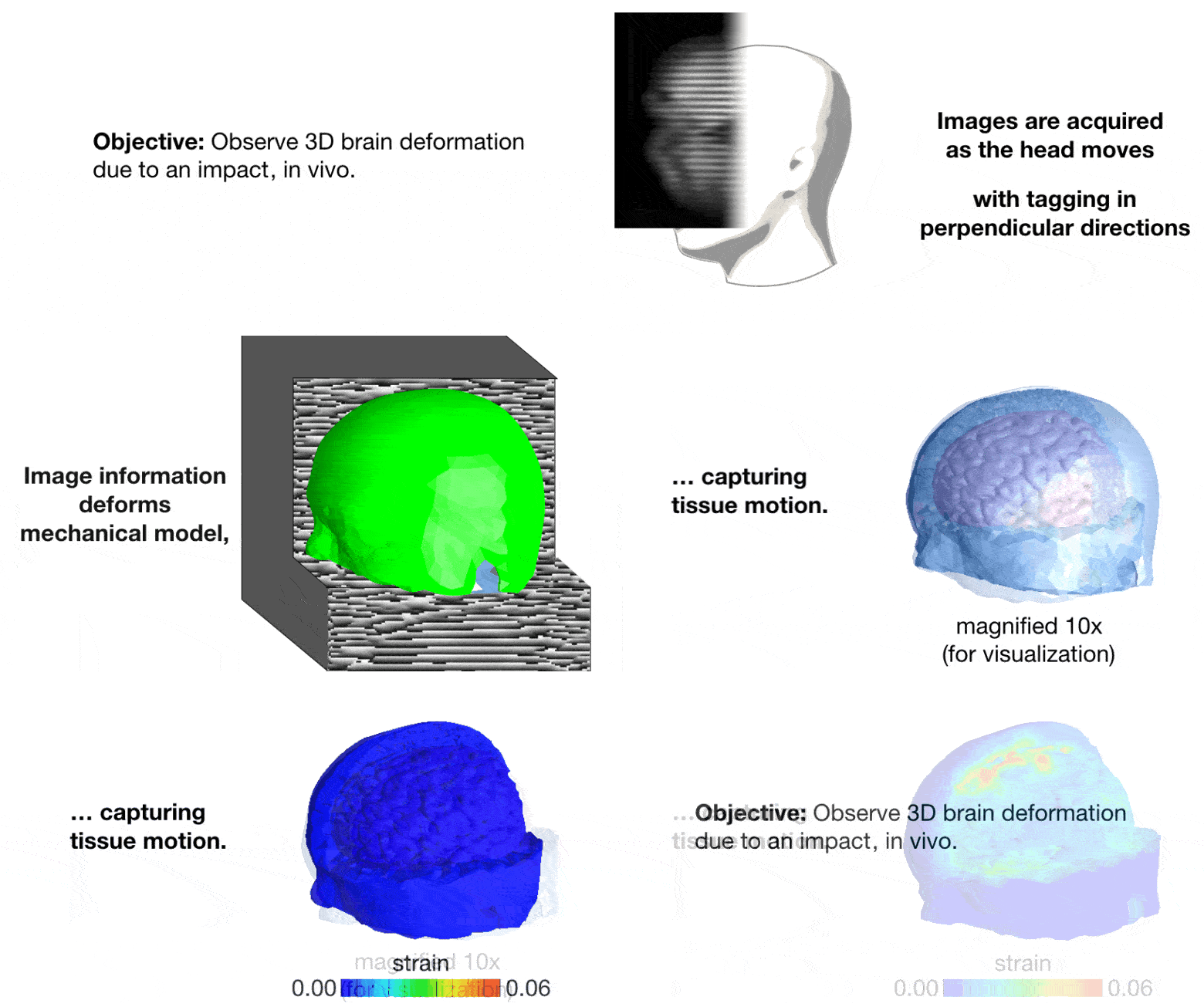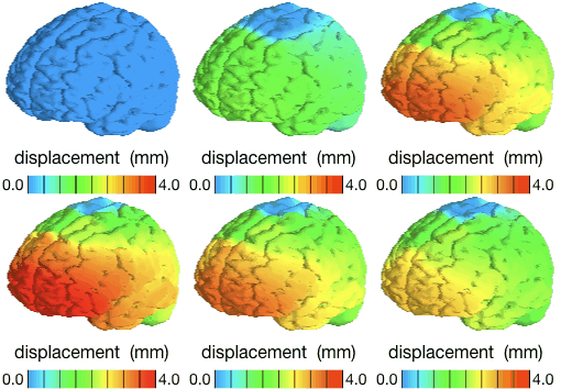
Measuring brain deformation enables researchers to better understand traumatic brain injuries and to develop experimentally verified computer models to design and evaluate preventive strategies. Although motion can be observed using medical imaging, quantifying fast tissue deformation based on inertially-induced motion is challenging due to the potential effects of noise and blurring, and the three-dimensional nature of motion. We present a technique, harmonic phase analysis with finite elements (HARP-FE), to estimate tissue deformation by combining multiple views of tagged magnetic resonance imaging and a biomechanical model of the brain. Spatial interpolation and a band pass filter are used to extract a volume containing the harmonic phase of a set of tagged images. These phase data are used to generate “pseudo-forces” that deform a finite-element model, causing the images to be registered following a mechanically feasible path. HARP-FE was calibrated using simulated brain deformation and data from an experimental phantom. The method was demonstrated human volunteers during head rotation (rotational acceleration) and neck extension (predominantly linear acceleration). Our results indicate that this method can quantify small brain deformations that occur during events typical of everyday activities. In particular, it can be used in vivo to capture the behavior of the intact, living human brain. We also show that the method delivers accurate estimations when some imaging information is corrupted. The combination of sensitivity to small deformations and robustness to practical imaging challenges will facilitate future studies of brain behavior under a larger range of conditions, and promises to enable measurements of motion in other organs as well.

