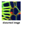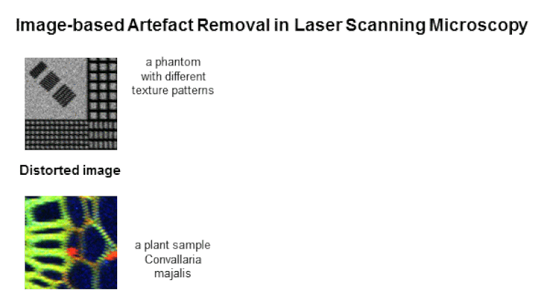
Advances in laser scanning microscopy (LSM) have greatly extended its applicability in cancer imaging not only to observe dynamic biological processes, but also to quantitatively measure them. The fast acquisition, with high spatial and temporal resolution and extended field of view enables scanning of larger areas of the specimen, and holds the promise of applying quantitative biology. In practice, however, many artefacts compromise image quality: the motion of the specimen due to cardiac, respiratory and other muscle contractions of the specimen, and the spatial image inconsistency (jaggedness) due to the varying speeds of the microscope laser. In this paper, we present a framework for efficient removal of jaggedness artefacts by a combination of two image-processing methods. First our framework compensates for the local displacements caused by the unknown, variable motion of the laser, and removes geometric distortion apparent in the acquired images. Second a fast local filtering procedure reduces the level of noise and improves the overall signal-to-noise ratio of the images. Our framework was quantitatively evaluated using phantom data from different microscope systems to show the capability of restoring corrupted images purely based on information from the acquired data. Generalisability of our framework was also demonstrated on cancer imaging applications of tumour vasculature growth, resulting in substantial improvement over other state-of-the-art microscopy acquisition methods. Furthermore, our framework could render formerly unusable data open to quantitative and correlative imaging analysis and would enable analysis of data that otherwise would have had to be discarded by biologists.Advances in laser scanning microscopy (LSM) have greatly extended its applicability in cancer imaging not only to observe dynamic biological processes, but also to quantitatively measure them. The fast acquisition, with high spatial and temporal resolution and extended field of view enables scanning of larger areas of the specimen, and holds the promise of applying quantitative biology. In practice, however, many artefacts compromise image quality: the motion of the specimen due to cardiac, respiratory and other muscle contractions of the specimen, and the spatial image inconsistency (jaggedness) due to the varying speeds of the microscope laser. In this paper, we present a framework for efficient removal of jaggedness artefacts by a combination of two image-processing methods. First our framework compensates for the local displacements caused by the unknown, variable motion of the laser, and removes geometric distortion apparent in the acquired images. Second a fast local filtering procedure reduces the level of noise and improves the overall signal-to-noise ratio of the images. Our framework was quantitatively evaluated using phantom data from different microscope systems to show the capability of restoring corrupted images purely based on information from the acquired data. Generalisability of our framework was also demonstrated on cancer imaging applications of tumour vasculature growth, resulting in substantial improvement over other state-of-the-art microscopy acquisition methods. Furthermore, our framework could render formerly unusable data open to quantitative and correlative imaging analysis and would enable analysis of data that otherwise would have had to be discarded by biologists.