
Time-of-flight (TOF) positron-emission tomography (PET) has increasingly been adopted as an important quantitative imaging tool in basic and clinical applications. Improving image quality via advanced image reconstruction methods remains an area of active interest. In particular, it is of practical significance to investigate image reconstruction from low-count TOF-PET data for shortening imaging time, increasing spatial-temporal coverage, enabling fast dynamic imaging, and/or reducing imaging dose.
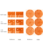
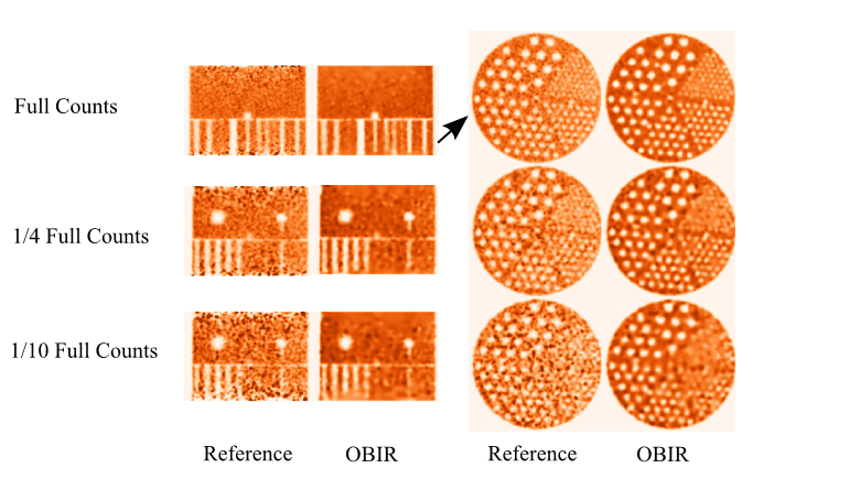
In this work, we investigate an optimization-based image reconstruction (OBIR) method with an emphasis on enabling image reconstruction from low-count data in TOF PET. In the OBIR method, we formulate TOF-PET reconstruction as a convex, non-smooth optimization problem composed of a Kullback-Leibler data divergence and an image-total-variation constraint; and we tailor the generic Chambolle-Pock primal-dual algorithm to reconstruct the image through solve the specified optimization problem. An advanced clinical TOF-PET scanner is used to collect both physical phantom and human subject data at full-count levels employed in current applications. Low-count data sets are extracted from the full-count data. We apply the OBIR method to reconstructing TOF-PET images from the full- and low-count data.
Using visual inspection and quantitative metrics, we evaluate spatial/contrast resolution, signal-to-noise ratio (SNR), and texture in images reconstructed by the OBIR method. The results indicate the investigated OBIR method has the potential to yield images with enhanced spatial/contrast resolution, increased SNR, minimized false lesion signals, and increased axial volume coverage over the images reconstructed (especially from low-count data) using the algorithm employed in the clinical scanner. The aforementioned image-quality enhancements could potentially be exploited for shortening imaging time, increasing spatial-temporal coverage, enabling fast dynamic imaging, and/or reducing imaging dose.