microbubbles

Contrast-free detection of focused ultrasound-induced blood-brain barrier opening using diffusion tensor imaging

Author(s)3: Maria Eleni Karakatsani, Antonios Pouliopoulos, Michael Liu, Sachin R. Jambawalikar, Elisa E. Konofagou
Contrast-free detection of focused ultrasound-induced blood-brain barrier opening using diffusion tensor imaging 170 177 IEEE Transactions on Biomedical Engineering (TBME)
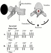
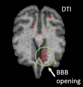
Karakatsani et al. report a contrast-free method to detect focused ultrasound (FUS)-induced blood-brain barrier (BBB) opening using diffusion tensor imaging (DTI). The localized BBB opening was hypothesized to result to a transient change in the diffusion pattern of water molecules and was tested in a non-human primate model, which closely resembled the human anatomy. They found that fractional anisotropy within the targeted area increased by 82% after the procedure, showing that DTI can confirm BBB opening without the use of contrast agents, increasing the safety of the methodology since contrast does not have to cross into the brain parenchyma. read more

Enhanced detection of bubble emissions through the intact spine for monitoring ultrasound-mediated blood-spinal cord barrier opening

Author(s)3: Stecia-Marie Fletcher, Natalia Ogrodnik, Meaghan O’Reilly
Enhanced detection of bubble emissions through the intact spine for monitoring ultrasound-mediated blood-spinal cord barrier opening 170 177 IEEE Transactions on Biomedical Engineering (TBME)
Short burst, phase keying (SBPK) is a clinically relevant pulse scheme for focused ultrasound delivery to the spinal canal. We demonstrate that emissions from microbubbles exposed to these pulses can be detected through ex vivo human vertebrae, and that SBPK exposures can open the blood-spinal cord barrier (BSCB) in rats. Microbubbles were sonicated through ex vivo vertebrae and emissions were analyzed using short time Fourier analysis. Pulses were modified to include pulse inversion, which enhanced detection of acoustic signatures. In rats, opening was confirmed by MRI enhancement. Detection of the subharmonic was linked to tissue damage observed at histology. read more