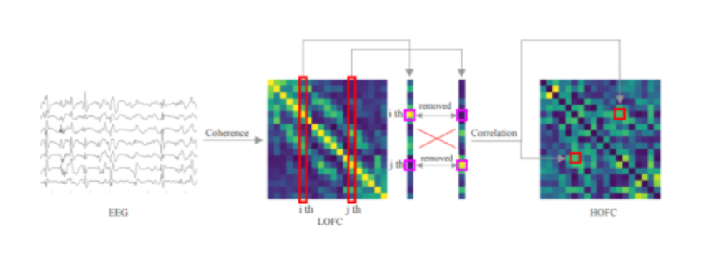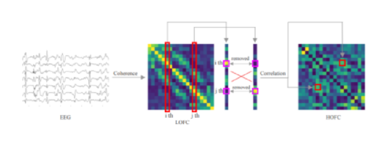
Previous studies made progress in the early diagnosis of Alzheimer’s disease (AD) using electroencephalography (EEG) without considering EEG connectivity. To fill this gap, we explored significant differences between early AD patients and controls based on frequency domain and spatial properties using functional connectivity in mild cognitive impairment (MCI) and mild AD datasets. Four global metrics, network resilience, connection-level metrics and node versatility were used to distinguish between controls and patients. The results show that the main frequency bands that are different between MCI patients and controls are the θ and low α bands, and the differently affected brain areas are the frontal, left temporal and parietal areas. Compared to MCI patients, in patients with mild AD, the main frequency bands that are different are the low and high α bands, and the main differently affected brain region is a larger right temporal area. Four LOFC bands were used as input to train the ResNet-18 model. For the MCI dataset, the average accuracy of 20 runs was 93.42% and the best accuracy was 98.33%, while for the mild AD dataset, the average accuracy was 98.54% and the best accuracy was 100%. To determine the timing of early treatment and discovering the susceptible patients, and to slow the progression of the disease, we assume that the occurrence of MCI and mild AD and their progression to more serious AD and dementia could be inferred by analyzing the topological structure of the brain network generated by EEG. Our findings provide a novel solution for connectome-based biomarker analysis to improve personalized medicine.

