A wearable brain scanner developed in the U.K. could take neuroscientists across the next big frontier in health care and help them to better understand the inner workings of a living brain
Today, the World Health Organization (WHO) reports that one in four people are affected by mental or neurological disorders, 50 m suffer from epilepsy, and 50 m from Alzheimer’s disease. According to the Journal of Neurosurgery, 69 m globally have some type of traumatic brain injury. With these numbers rising daily, there is an urgent need to understand what is happening inside the brain. Brain scanning technology has advanced dramatically in recent decades, but it still faces significant limitations.
“There are two main types of information we can get from a brain scanner,” says Dr. Niall Holmes, research fellow at the University of Nottingham, Nottingham, U.K., and co-founder of Cerca Magnetics Ltd. (Figure 1). Magnetic resonance imaging (MRI) and X-ray CT machines show brain structure but do not provide information about brain function, including the timings and firings of neurons. Functional scanners such as electroencephalography (EEG) systems show the timings of neuronal activity but provide only limited information about where in the brain it is happening. “It can’t show us exactly where an epileptic center is, for example,” he adds. “Few technologies can do both structure and function, so that is what we wanted to develop.”
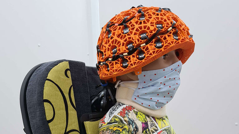
In his research, Holmes’ uses quantum technologies to create wearable devices for magnetoencephalography (MEG), a functional neuroimaging technique that measures magnetic fields generated by neuronal currents. By combining quantum magnetic field sensors and novel magnetic shielding to screen out interference, he and his colleagues have been able to perform the first MEG recordings while subjects perform significant physical movements. This opens up a wealth of possibilities including scanning children and patients with movement disorders, and could enable neuroscientists to incorporate technologies such as virtual reality (VR) to provide a more immersive environment.

In from the cold
For Holmes, the journey into brain imaging began with a personal moment when studying for his A levels, the U.K. equivalent of American Advanced Placement courses. “I was around hospital environments with my father who was ill, and I saw the radiotherapy technology and other equipment, so I was interested to see how physics fed into medicine,” he says. “Then I discovered you could do medical physics courses and chose one at Nottingham University, the home of MRI.”
Nottingham alumnus Sir Peter Mansfield, who was awarded the Nobel Prize for medicine in 2003, developed calculation methods in the 1970s that contributed to the development of MRI. Holmes, who finished his Ph.D. in 2019 and also completed an internship at the Sir Peter Mansfield Imaging Center, is now helping the university stay at the cutting edge of scanner development.
“MEG is a very promising way to improve brain imaging, but it is incredibly difficult,” remarks Holmes. “Magnetic fields generated by the brain are a hundred billion times smaller than the earth’s field, so it is hard to measure them.”
Previously, systems deployed cryogenic sensors—superconducting quantum interference devices (SQUIDs)—that needed to be cooled to very low temperatures. To restrict movement, patients were fixed inside a scanner, which was expensive to run due to the high volume of liquid helium it consumed on a daily basis. The recent surge in quantum technologies has, however, enabled many of these challenges to be overcome. Using highly sensitive quantum magnetometers, Holmes and his team have been able to make a functioning wearable MEG system.
Into the quantum realm
It is often said that anyone who claims to understand quantum physics does not understand it at all, as it involves the study of energy and matter at the most fundamental level, focusing on the behavior of subatomic particles—the building blocks of all things. Though it is riddled with complexity, Holmes is nevertheless able to describe the operation of quantum magnetometers in a relatively simple way.
“Each sensor contains a small cloud of rubidium atoms, which have the quantum mechanical property of spin,” he explains. “Imagine each atom as a compass needle pointing in a different direction. When there is no magnetic field and you shine a laser through the cloud, the atoms align with the direction of the laser, and we can measure the laser’s intensity with a sensor.”
“Magnetic fields then disturb the compass needles, attracting them, so they fall out of alignment with the laser, but the atoms want to align with the laser, so momentum is transferred from the laser to the atoms to achieve realignment,” Holmes adds. “The measurement of the laser’s intensity decreases during this process, and we use that as an indicator of the magnetic field experienced by the cloud that is coming from the brain.”
Developing quantum sensors that are robust enough to deliver readable results in a therapeutic setting is a major technical challenge, but it is one that the team at Nottingham and at Cerca has overcome with the help of U.S. company QuSpin, which designs and builds the sensors. “As well as challenges with quantum technologies, there is also the issue of the order of magnitude between the signal you want to detect and the signals that distort the magnetic field in the sensors,” Holmes observes. “A lot of my work focuses on magnetic shielding and interference rejection. Even a bracelet or an earring can mask the brain’s activity.”

Room to move
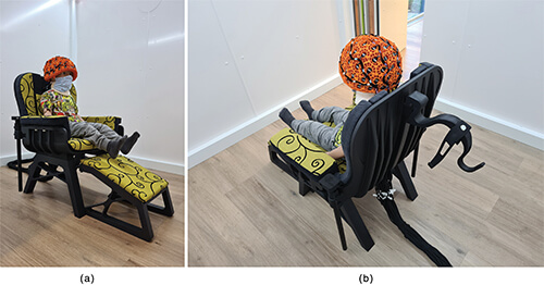
The MEG is a wearable device, but the sensitivity to outside disruption means that it cannot be used everywhere or with subjects walking freely around their environment. Signals from the quantum sensors are easily distorted, so they must be used in a room that is heavily shielded against external magnetic fields [Figures 2 (a) and (b)].
The solution Holmes has developed is similar to a Faraday cage—a tried and tested version of an enclosure used to block electromagnetic fields—that uses a mesh of conductive material to absorb incoming signals. These cages were invented by pioneer Michael Faraday back in 1836, but for the MEG to work effectively a more complex magnetically shielded room (MSR) is required.
“I worked on additional magnetic shielding with electromagnetic coils to cancel out the magnetic field,” Holmes says. “It works in a similar way to noise-canceling headphones or a humbucking coil on a guitar, which use phase cancellation to reduce interfering signals and get a cleaner sound.”
The MSR cuts out the earth’s magnetic field, though a spatially varying remnant field remains. Any movement of the quantum sensors through the remnant field can overload the inputs and make the resulting data unusable. Holmes, therefore, developed a system of six large bi-planar electromagnetic coils that cancel the remnant field inside the MSR, which allows subjects some degree of movement while being scanned.
“The need for MSRs means that the device is not really portable, though it is wearable,” he adds. “It cannot be taken out of the rooms, which are large spaces and, therefore, expensive to build. What we have to do now is make the rooms cheaper to construct. We also need to work on making the sensing technology better so that we can use little or, ultimately, no shielding. If we can do that, the technology could be installed in a doctor’s surgery.”
Diving deep into the brain
Research toward those goals is already progressing rapidly. The University of Nottingham team is collaborating with U.K.-based specialist manufacturer magnetic shields. Furthermore, there has been enthusiastic support for the technology from neuroscientists, including a team at Great Ormond Street Hospital, London, U.K., that is investigating epilepsy.
Cerca has already received the first order for a MEG system that will be dedicated to clinical use. It will be used by a U.K. research organization to study epilepsy and, although it will not be used to make clinical decisions, it will enable research trials that will shed light on the causes and characteristics of the condition.
Another potentially groundbreaking application of the system is in the field of autism research. The Hospital for Sick Children, Toronto, ON, Canada, will use MEG to study the brains of autistic children to explore a condition that is more frequently diagnosed than ever before but is poorly understood. Research from Newcastle University, the University of Cambridge’s Department of Psychiatry, and Maastricht University recently showed that around one in 57 children in the U.K. is on the autistic spectrum, significantly higher than previously reported.
What is clear is that a wearable MEG system could be used to investigate any neurological condition from concussion to Parkinson’s disease. “MRI scanners are not very comfortable for an adult, let alone a child,” says Holmes. “Infants often need sedation so they don’t move. With our technology, we can scan across lifespan because it is so comfortable and the data is so good. Neuroscientists know a lot about the differences between ‘healthy’ brains and brains with certain conditions, but we don’t know much about how the brain changes over time and when developmental differences start.” The MEG system has already been used successfully to scan children as young as two years old.
“With a movement disorder, for example, we need to activate the brain networks that are not working properly, so we need people to engage in activities rather than just lying down and watching a screen,” Holmes remarks. “And we can combine it with other technologies like VR to create different stimuli, or we can scan mother and baby interacting and compare it to a stranger and baby.”
“We want to scan in as natural an environment as possible,” he adds. “We are looking at scanning newborn babies and people making bigger movements, exploring the space in the room. We need to balance the needs of clinical centers with the physics element, but we have been able to communicate clearly what we want to do and there is a lot of interest out there.”
Although the technology is at a very early stage—there are only three systems in the U.K.—there is a groundswell of support for the research. As it improves, becomes cheaper, and enables scanning for a bigger range of movement, MEG could soon become the biggest story in brain imaging.


