Atlases of anatomy have long been a mainstay for visualizing and identifying features of the human body [1]. Many are constructed of idealized illustrations rendered so that structures are presented as three-dimensional (3-D) pictures. Others have employed photographs of actual dissections. Still others are composed of collections of artist renderings of organs or areas of interest. All rely on a basically two-dimensional (2-D) graphic display to depict and allow for a better understanding of a complicated 3-D structure.
In the life sciences, the relationships between biological structure and function have been central to the understanding of health and disease throughout recorded history. For many centuries, the illustrations of anatomy originally created during the medieval period formed the basis for the study of medicine. An intimate understanding of 3-D biological structure and its implications for therapy remains essential for medical specialties such as surgery, neurology, and radiology and a learning challenge for all health professionals. But a fundamental limitation to the teaching, learning, and understanding of 3-D structure is that most pictures and illustrations reproduced in books or as plain film radiographs are fundamentally 2-D in nature. The usual attempt to convey 3-D content through a series of related 2-D images forces upon the viewer an exercise in mental model construction.
Medical Applications of Digital Images
The practice of all medicine is 3-D. The practice of neurology, reconstructive surgery, neurosurgery, orthopedics, radiology, and radiation therapy is dependent on 3-D understanding and imaging. Stereotactic guidance for neurosurgical procedures using 3-D imaging data is no longer just a dream. Treatment of bone abnormalities often includes preoperative 3-D imaging. The images are intended to convey a clear anatomic model to the physician as well as to build prosthetic devices for implantation and reconstruction that fit the particular patient [2]. Such data have also been used to guide the actual robotic milling of bone and joint surfaces in vivo.
Planning Panel on Electronic Imaging
Early in 1989, under the direction of the Board of Regents of the U.S. National Library of Medicine (NLM), an ad hoc planning panel was convened to provide the library with in-depth guidance as to its proper role in the rapidly changing field of digital imaging. The panel recognized that much of our understanding of complicated health and disease processes actually lies in images, not text. Over the centuries, we have been using text to describe our view of body systems, organs, and molecules because of the difficulty and expense of creating and distributing the images. The panel made the following recommendation [3]:
The NLM should undertake a first project, building a digital image library of volumetric data representing a complete normal adult human male and female. This “Visible Human Project” would include digitized photographic images from cryosectioning, digital images derived from computerized tomography, and digital magnetic resonance images of cadavers.
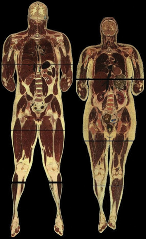
The details of how the images that make up the Visible Human data sets were captured are described elsewhere [4]. The male data set, 15 gigabytes released in November 1994, contains 1,871 digital axial anatomical images obtained at 1.0-millimeter intervals and associated computerized tomography (CT) and magnetic resonance imaging (MRI) images. The female data set, 39 gigabytes released a year later in November 1995, contains 5,189 digital axial anatomical images obtained at 0.33-millimeter intervals and associated CT and MRI images generally obtained at 1.0-millimeter intervals. Figure 1 illustrates the male and female anatomical data.
Applications of the Data Sets
The Visible Human Project data sets are available to researchers from academia and industry or to individuals who are interested in using the digital image data produced by this project through a nonfinancial licensing agreement with the NLM. This approach makes it possible for interested parties to help the NLM reach a consensus on the collection, maintenance, and distribution processes for digital image libraries that will be mutually beneficial. License holders may obtain the data sets by downloading them via the Internet using the File Transfer Protocol.
The data sets are being applied to a wide range of educational, diagnostic, treatment planning, virtual reality, artistic, mathematical, and industrial uses by over 3,500 licensees in 64 countries. In health care and health education, they are used as a normal reference and as an aid in the diagnostic process. Programs have been developed to educate patients about the need for and purpose of surgery and other medical procedures, as well as to permit physicians to plan surgery and radiation therapy. The images from the Visible Human data sets are used in several prototype virtual reality surgical simulators, including virtual colonoscopy. Educational materials that make use of the Visible Human data sets are being used by students from kindergarten to practicing healthcare professionals, provide a starting point for medical illustrators, and serve as a common source of images for the development and testing of rendering algorithms.
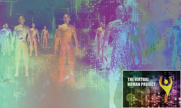
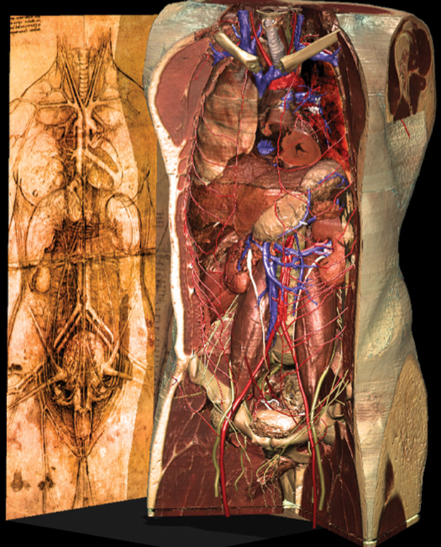
In nonhealthcare applications, the data sets form the basis of interactive games to entertain as well as to educate. Automobile manufacturers now include passenger injury models based on Visible Human data in their vehicle crash simulation testing. Engineers and physicists have created models to quantify human exposure to various forms of electromagnetic radiation. The data provided by the Visible Human data sets have been used by mathematicians as an application for what were previously only theoretical mapping theories. Several artists have used the data sets as the basis for new multimedia art forms (Figure 2).
While originally conceived as a resource for teaching human anatomy, as 3-D imaging has become more widely available, the Visible Human data sets are finding use beyond anatomy in various medical specialties and, beyond medicine, in engineering, modeling, virtual reality, and art. More than 20 years later, the data sets continue to be licensed for use in health- and non-health-related applications.
References
- M. J. Ackerman, “The Visible Human Project,” Proc. IEEE, vol. 86, no. 3, pp. 504–511, Mar. 1998.
- A. M. DiGioia, B. Jaramaz, R. V. O’Toole, D. A. Simon, and T. Kanade, “Medical robotics and computer assisted surgery in orthopaedics,” in Interactive Technology and the New Paradigm for Health Care, K. S. Morgan, R. M. Satava, H. B. Sieburg, R. Masttheus, and J. P. Christensen, Eds. Amsterdam, The Netherlands: IOS Press, 1995, pp. 88–90.
- “Electronic imaging: Report of the Board of Regents,” Board of Regents, National Library of Medicine, National Institutes of Health, U.S. Department of Health and Human Services, NIH Publication 90–2197, 1990.
- V. Spitzer, M. J. Ackerman, A. L. Scherzinger, and D. Whitlock, “The Visible Human male: A technical report,” J. Amer. Med. Assoc., vol. 3, no. 2, pp. 118–130, 1996.