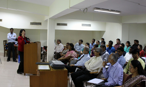
A two day interdisciplinary workshop on Achieving In situ Functional Histopathology (AifH 2015) was held in March at the Indian Institute of Technology (IIT) Kharagpur, India. The workshop showcased invited lectures from eminent academicians, clinicians and inventors in the field of medical imaging who are specifically working on technologies to understand pathology of tissues in situ. Coordinated by Dr. Jyotirmoy Chatterjee and Dr. Debdoot Sheet with extended cooperation from Dr. Pranab K. Dutta, Dr. Siddhartha Sen, Dr. Anirban Mukherjee and Dr. Manjunatha Mahadevappa from IIT Kharagpur, India, the workshop provided 30 young researchers with a platform to network and learn from experts in this multi-disciplinary area of translational research in computational and biomedical imaging.
The workshop started with Prof. Pranab K. Dutta from IIT Kharagpur, speaking on diffusion near-infrared imaging and diffusion tomography. Prof. Mousumi Pal and Prof. Ranjan Rashmi Paul from the Guru Nanak Institute of Dental Science and Research, Kolkata, followed with their talk on clinical challenges with in situ diagnosis of oral cancers and lesions. Prof. May D. Wang, and IEEE EMBS Distinguished Lecturer from the Georgia Institute of Technology and Emory University, Atlanta, GA, USA, spoke on biomedical imaging big data analytics for personalized care. Prof. Jyotirmoy Chatterjee from IIT Kharagpur, followed with his talk on multimodal imaging and analysis for in situ functional histology in advancing the understanding of systems pathology. Prof. Krishna Dalal from the All India Institute of Medical Sciences (AIIMS), New Delhi, presented her work using optical coherence tomography for evaluating healing efficacy of traditional Indian medicine in treating dermatological conditions. Prof. Sambuddha Ghosh from the North Bengal Medical College and Hospital in Darjeeling presented the success of large scale acceptance of fundus autofluorescence imaging among ophthalmologists for in situ functional investigation of retinal tissue health. Dr. S. R. Narahari from the Institute of Applied Dermatology, Kasaragod, Kerala, and Dr. B. Vijaya from JSS Medical College, Mysore, Karnataka, jointly spoke on their work in developing ayurvedic (traditional Indian medicine) practices for diagnosing and treating Psoriasis and evaluating its healing efficacy using dermatopathology, immunohistochemistry for integrative medicine. Dr. Chilakapati Murali Krishna from the Advanced Centre for Treatment, Research and Education in Cancer (ACTREC), Mumbai, shared his work on optical and spectral pathology using Raman spectroscopy. Dr. Sharmistha Paul from the West Bengal State Council of Science and Technology, Kolkata, spoke about her work on the surface plasmon resonance imaging tool for understanding biomolecular interactions in situ. Prof. Basu Dev Banerjee from the University College of Medical Sciences, New Delhi, presented his work on inflammatory pathway genes and organochlorine pesticide levels as the risk factor assessment for epithelial ovarian cancer. Dr. Rita Banerjee from the Ministry of Science and Technology, Government of India, New Delhi, provided insight about their initiatives with promoting multimodal imaging in the diagnosis of disease.
The first day’s program also featured four talks delivered by senior students about their research work, with Anji Anura from IIT Kharagpur, speaking about her work with exploring molecular pathology based markers for screening malignant potentiality of oral pre-cancers. Biswajoy Ghosh from IIT Kharagpur followed with his journey to achieve in situ optical biopsy. Sri Phani Krishna Karri from IIT Kharagpur spoke about his experiences with computational imaging meeting microscopy and Sushmita Dey from the Indian Institute of Engineering, Science and Technology (IIEST), Shibpur, Howrah, India, presented her work on multimodal characterization of oral exfoliative cytology in smokers for cancer risk assessment.
The second day of the workshop featured Prof. Amita Giri from the North Bengal Medical College in Darjeeling, speaking about problems in early diagnosis of oral pre-cancerous lesions. Prof. Bindu M. Kutty from the National Institute of Mental Health and Neurological Sciences (NIMHANS) in Bangalore spoke about implications of neuro-morphological imaging studies in understanding the cognitive reserve capacities of the brain. Prof. Pradeep K. Gupta from the Raja Ramanna Centre for Advanced Technology (RRCAT) in Indore followed with using optical spectroscopic and imaging techniques for biomedical diagnosis. Dr. Jay Sengupta from the Minneapolis Heart Institute at Abbott Northwestern Hospital, Minneapolis, USA spoke about methods to assess tissue characteristics in atrial fibrillation and catheter ablation. Prof. Provas Banerjee from the Hi-Tech Medical College and Hospital, Bhubaneswar, India shared his clinical experiences with using alternative medical practices for wound management and evaluating healing progression with in situ imaging like optical coherence tomography (OCT). Prof. Sanghamitra Sengupta from the University of Calcutta, Kolkata, India spoke about microbiome, intestinal integrity and disease pathogenesis. Dr. Raunak K. Das from the Vellore Institute of Technology (VIT) University, spoke about correlating microscopy and spectroscopy of oral tissues. Dr. Tandra Sarkar from the Apollo Hospital, in Kolkata spoke about clinical practices in molecular imaging for breast cancer detection. The technical program concluded with Dr. Debdoot Sheet from IIT Kharagpur speaking about hierarchical machine learning technologies for understanding tissue-energy interaction in ultrasonic and optical imaging for developing in situ histology technologies.
The workshop also featured two hands on learning sessions for the participants about biopsy tissue processing, bright-field and phase-contrast optical microscopy, ultrasonic imaging, and optical coherence tomography. The workshop concluded with a musical evening. This workshop was supported by the Science and Engineering Research Board (SERB) of the Department of Science and Technology, India the Indian Council for Medical Research (ICMR), India and the IEEE Engineering in Medicine and Biology Society.