Who teaches us more than the beasts of the earth, and makes us wiser than the fowls of heaven?
—Job 35:11
Medical science developed in tandem with the evolution of biological species and their associated diseases. Because of the close interaction between humans and other animals, even those in the wild, taking care of the former also means caring for the latter. Several scientific forerunners delved into animals’ anatomical and physiological secrets in their quest to better understand animal biology and functions, thereby laying the foundation for animal medicine. Here, I briefly explore the long and complex road that led to the current state of veterinary science and provide a few examples of its present standing. (Contributions from the ancient world and eastern countries are not considered, as they represent a different area of interest.)
The Forerunners
After a long and dark period of 1,000 years, between 400 and 1,400 CE, when science remained essentially stagnant, the modern European period (1400– 1800) slowly emerged from that prolonged slumber. Leonardo Da Vinci (1452–1519) was one of its earliest progenitors of this Renaissance—although, unfortunately, with relatively little scientific influence, mostly due to his obsession with secrecy [1].
Andreas Vesalius
One unquestionably outstanding early contributor to the ignition of this new intellectual flame, and a true bridge between the Middle Ages and modern history, was Andreas Vesalius (1514– 1564). Born in Brussels, Belgium, then part of the Holy Roman Empire, Vesalius came from a family of physicians and, following this tradition, studied medicine in Paris. Forced to leave before completing his degree when the empire declared war on France, he proceeded to the University of Louvain and thereafter moved to Padua, where he soon held the chair of surgery and anatomy.
Vesalius believed that surgery had to be grounded in anatomy. Unusual for the time, he always performed dissections to produce anatomical charts. In a small booklet on bloodletting (then a popular treatment for several illnesses), Vesalius showed clearly how anatomical dissection could be used to disprove fruitless speculations. In 1539, his supply of dissection material increased when the bodies of executed criminals became available to him. This is a marked departure from Galen (129–210 CE), whose anatomy was plagued with errors and misconceptions ( although some of his descriptions were acceptable and some of his teachings useful).
In 1543, Vesalius published De Humani Corporis Fabrica, the first text based largely on human dissection, transforming anatomy into a subject that relied on observations. It represented a considerable effort and may have exhausted him because, after finishing the book, Vesalius left anatomical research and teaching and took up medical practice, serving the imperial court of Emperor Charles V and later Charles’ son, Philip II of Spain. In 1564, he made an unexplained trip to the Holy Land but died on the Greek island of Zakynthos, perhaps felled by a contagious disease or poor sanitary conditions on the ship. No document concerning his death has been found on the island, and, sadly, no one has yet been able to find the site of his burial. However, he is highly recognized and honored in Zakynthos, where a statue of Vesalius was erected [3], [27], [28].
William Harvey
Born 14 years after Vesalius’s death, William Harvey (1578–1657) was the first to correctly describe the circulation of blood and demonstrate that arteries and veins form a complete circuit, starting at and leading back to the heart, while the heart’s regular contractions drive the flow of blood throughout the body. In 1593, at just 15, Harvey enrolled as a medical student at the University of Cambridge, England, supported by a six-year scholarship. He had the opportunity to spend some time at universities in France, Germany, and Italy, thus improving his knowledge of science and medicine. Then in 1599, when only 21, he enrolled at the University of Padua in Italy, where Hieronymus Fabricius (1537–1619), a skilled anatomist and surgeon, became his dear and respected teacher. (Fabricius had discovered valves in human veins in 1574, although he did not publish the discovery until 1603.) An outstanding student, Harvey graduated in 1602.
On his return to England, the University of Cambridge awarded him a Doctor of Medicine degree, adding to the one he already had from Padua, and he then moved to London to work as a physician, joining the College of Physicians in 1604. He became a fellow of the college in 1607 and head physician at Saint Bartholomew Hospital. By the time he was 40, Harvey had won recognition as London’s best surgeon and was appointed physician to King James in 1618 and later to King Charles. Quite a brilliant career. In 1628, Harvey published his masterpiece, Anatomical Studies on the Motion of the Heart and Blood in Animals or, for short, De Motu Cordis [4], based on animal dissection.
Harvey’s knowledge came from his own observations of blood flowing through the veins and arteries of several species of living animals, which he cut open (obviously without anesthetics). Ironically, in correcting Galen’s mistakes, Harvey ran into trouble with other European physicians, who made great use of Galen’s worthless bloodletting methods; his own medical practice declined because of strong criticism from these quacks. In fact, even almost 300 years after Harvey’s death, Galen’s bloodletting methods were still being used in many places all over the world! Harvey refuted a number of then-standard beliefs about how the heart and blood system worked, clearly establishing the following ten essential truths, valid for humans, other mammals, and even other nonmammalian species.
- The blood in arteries and veins is all of the same origin, not manufactured in different parts of the body.
- The blood that is sent through the arteries to the tissues is not consumed there.
- The circulation mechanism is designed for the movement of liquid, not air, and the blood on the right side is still blood.
- The heart is the source of blood movement, not the liver.
- The heart contracts at the same time that the pulse is felt.
- The ventricles squeeze blood into the aorta and pulmonary artery.
- The pulse is not produced by the arteries pulling blood in but by blood being pushed by the heart into the arteries, enlarging them.
- There are no vessels in the heart’s septum, and all blood in the right ventricle goes to the lungs and then through the pulmonary veins to the left ventricle.
- All blood in the left ventricle is sent into the arteries, routed by the smaller veins into the venae cavae, and then to the right ventricle again, thereby completing the circulation.
- There is no to-and-fro movement of blood in the veins but a constant flow of blood to the heart.
William Harvey died in London, at 79; his grave can be found in the village of Hempstead, in the English county of Essex [5].
Stephen Hales
At 19, Stephen Hales (1677–1761, born in Beckesbourne, Kent, England) entered the University of Cambridge (the same university Harvey attended), obtaining the Master of Arts degree in 1703. He studied comparative anatomy, dissecting frogs, dogs, and other animals (providing good early studies for veterinarian activity). In 1710, Hales was made a perpetual curate at Teddington in Middlesex, where he was ordained in the ministry, receiving in 1711 the Bachelor of Divinity degree. Teddington became his home for the remainder of his life. In 1718, Hales was elected a fellow of the Royal Society and also became rector of Porlock, Somerset, a post he held alongside the curacy of Teddington. In 1723, he was elected rector of Farringdon, Hampshire (which he held alongside Teddington).
Hales’s fame as a scientist grew from 1718 onward; by the middle of the 18th century, he had achieved an international reputation. Hales was one of the eight foreign members of the Royal Academy of Sciences, Paris, and was elected a member of the Academy of Sciences of Bologna. He was made a Doctor of Divinity by Oxford University in 1733. Hales died at 84 in January 1761 after a short illness. At his own request, he was buried under the tower of the church where he had worked for so many years.
Hales began his work on animal physiology with William Stukeley (1687–1765) [6], who initially studied medicine (also at Cambridge) and later in life was ordained vicar of All Saints Church in Stamford, Lincolnshire. Both men performed a wide range of studies, including casts of the trachea and bronchial trees of dogs using molten lead and measuring the water lost due to breathing. Most famously, Hales made measurements of blood pressure in several animal species by inserting fine tubes into arteries and calculating the height to which the column of blood rose. Hales also described the effects of hemorrhage and hemorrhagic shock by progressive exsanguination of animals and the accompanying measurement of blood pressure. In a mare, he observed that, as death approached, the animal fell into cold and clammy sweats.
In addition, Hales took wax casts of the ventricle of the heart and estimated how much blood was pumped by the heart; correctly described the roles of the mitral and aortic valves during systole and diastole; explained the pulsations of arteries in terms of their elasticity; and attributed the resistance to blood flow to friction due to the passage of blood through small blood vessels. His Statical Essays was published in 1727; its famous first paragraph clearly and precisely describes arterial blood pulsations. There were two other editions, published in 1731 and in 1733 [7]. These experiments introduced the vertical open-end manometer, measuring an oscillating blood pressure in inches of blood (which we now know was really mean pressure) [8].
Jean-Louis-Marie Poiseuille
A French physician and physiologist, Jean- Louis-Marie Poiseuille (1799–1869) formulated a mathematical expression for the flow rate of the laminar (nonturbulent) movement of fluids in circular tubes. Discovered independently by Gotthilf Heinrich Ludwig Hagen (1797–1884), a German hydraulic engineer, this relation is also known as the Hagen–Poiseuille equation. Poiseuille received his medical degree in 1828 and established his practice in Paris. His interest in the circulation of blood led him to conduct a series of animal experiments and, thereafter, experiments on the flow of liquids in narrow tubes, from which he deduced the law that bears his name. This equation states that the flow rate is determined by the viscosity of the fluid, the drop in pressure along the tube, and the tube diameter. Poiseuille was also the first to use the mercury manometer to measure blood pressure and tried to prevent coagulation during the measurements [9].
Birth of Veterinary Medicine
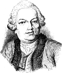
Except for some references to animal healers that go back to about 3,000 BCE in Asia Minor, the 1761 founding of the veterinary school in Lyon, France, by Claude Bourgelat (1712– 1779) marks the true beginning of the veterinary profession (Figure 1, right – Image courtesy of Wikimedia Commons.). Thereafter, in 1785, the Odiham Agricultural Society in England promoted the study of farriery based on rational scientific principles (farriery being the best way of producing and fitting horseshoes, as horses represented an essential mode of transportation). Out of the society’s deliberations came the establishment of the London Veterinary College in 1791, a professional group dedicated to animal medicine. Initially, the veterinary profession was centered on the horse, and this remained the focus for many years, influenced by the needs of the military.
Over time, the interests of the profession spread to cattle and other livestock, then to dogs, and now to companion and exotic animals. But who thought in those early days to clinically measure, say, arterial blood pressure in animals (even though some research had been conducted on the topic, as mentioned in [10])? The history of clinical medicine reflects both the complex nature of the relationship between physicians and patients and the slow evolution of knowledge in the biomedical sciences, specifically on the application of science to the control and management of human disease. Its extension to the veterinary area, first to domestic animals and much more recently to wild species, required a long time and a profound evolution of concepts and ideas [11].
After his father’s death and still an adolescent, Bourgelat was possessed of very scant means to proceed in life. Between 1724 and 1729, he served in the company of the musketeers. Then, we find him in Lyon, his native town, where he had a law firm from 1733 to 1740 that allowed him to become acquainted with the local bourgeoisie and ultimately realize his professional ambitions. In 1740, at age 28, Bourgelat obtained the position of écuyer (esquire) at the Académie d’Équitation de Lyon, holding this position until 1765. In 1744, he published a treatise on horse riding that was well received in Europe because of its novel approach and, in particular, because it called attention to previous errors in equine biomechanics [12]. Bourgelat decided to devote himself to this area and requested the help of two human specialists, Claude Pouteau and Jean-Baptiste Charmetton, both professors of surgery in Lyon, who enthusiastically accepted the invitation. They soon realized that there were similarities and differences between the human and animal machines and saw a clear opportunity to create medical treatment for animals.
In 1761, Bourgelat was authorized and given the necessary funds by the King of France, Louis XV, to found his veterinary school in Lyon. The school officially opened in February 1762. Another school was later established in Paris, with Bourgelat appointed as director and general inspector of the veterinary schools of the kingdom. These schools accepted students from other European countries, such as Switzerland, England, Sweden, Denmark, Germany, and Italy. Other schools were started by these former students, as offprings of the initial ones.
Thus, Claude Bourgelat became the true and undisputable father of veterinary medicine. One of his concepts, still valid, states that studying human medicine is useful to the study of medicine for horses, and vice versa [13], an idea later confirmed by several authors [14]–[18]. In addition, the influence of Carl Ludwig, his kymograph, and his school must be recognized, along with the superb studies of the horse made by Chauveau and Marey [19]–[22].
Discussion
Veterinary medicine has long since come of age and is now a well-established and fully recognized science and profession dealing with the prevention, diagnosis, and treatment of disease, disorders, and injury in animals, domesticated and wild, covering a wide range of conditions affecting many different species. Veterinary science helps human health directly through the monitoring and control of zoonotic diseases and food safety and indirectly through human applications of basic animal research. It also helps to maintain the food supply through livestock health monitoring and treatment. Veterinary scientists often collaborate with epidemiologists and other health or natural scientists [23].
It is now known that optimal movement and mobility can significantly impact the physical and mental health of animals. Canine rehabilitation has moved to the forefront of modern veterinary medicine with the advent of the American College of Veterinary Sports Medicine and Rehabilitation. Mechanical appliances are being used to improve the mobility and functionality of impaired patients, no longer the purview of human medicine alone. Veterinarians already have a history of creating assistive devices from materials at hand, using everything from plywood to low-temperature thermoplastics and aluminum rods to polyvinyl chloride pipes. As our understanding of the intricacies of quadruped mobility and biomechanics has grown, so have the variety and sophistication of these devices. Now they incorporate veterinary-specific hinges, composite plastics, titanium, carbon fiber, and specialty foam liners, making direct use of materials science products. Veterinary orthotics and prosthetics is evolving into a new specialty.
Orthoses (braces) are any medical device attached to the body to support, align, position, immobilize, prevent, or correct deformity; assist weak muscles; or control and improve function. They are not a replacement for surgery but, rather, complementary. Orthoses can be used as preoperative or postoperative solutions or even as alternatives to surgery. In cases in which surgery must be delayed, they can provide interim support, protect a limb, and minimize disuse atrophy. Postoperatively, orthoses can provide a safe, effective, and dynamic alternative to casting. Orthoses can also be used when surgery is not an option, such as in patients who are poor candidates for anesthesia, have comorbidities, or are advanced in age, or if finances are an issue for the individual.
The structural consequences of a missing limb or limb segment are now being recognized, in part through the efforts of rehabilitation therapists who understand the biomechanics of locomotion. Re-establishing a quadruped structure should be the goal whenever possible. Prostheses, like orthoses, are readily accepted by veterinary patients with congenital limb deformities as well as by those requiring an elective-level amputation. In human medicine, amputation at the hip for a catastrophic ankle injury would be unthinkable, but elective-level amputation is a recent development in veterinary medicine. It can improve quality of life and functional independence, preventing premature decisions to euthanize. Patients can return to an active lifestyle that curtails obesity and associated problems. Biomechanics can be improved, decreasing secondary or compensatory pain [24].
A couple of examples better clarify the wide spectrum covered by veterinary science. Brucellosis is caused by gramnegative coccobacilli of the genus Brucella. In livestock, the disease results in significant economic losses due to reproductive impairment caused by abortion, stillborn or weak calves, neonatal mortality, and infertility. In humans, the Brucella infection causes a febrile disease that may be associated with a broad spectrum of symptoms; in some cases, it may be fatal [25].
As established in the foundational principles of veterinary science, research and the care of wild species are also part of its concerns. For example, the tuco-tuco (Ctenomys affinitas knighti) is a subterranean rodent inhabiting a semiarid area in northwestern Argentina. Although the animals live in underground burrows where environmental cycles are attenuated, they display robust, 24-hour locomotor activity rhythms that are synchronized by light/dark cycles, both in laboratory and field conditions. The underground environment also poses energy- related challenges (e.g., the demands of digging, hypoxia, high humidity, and reduced food availability); these have motivated thermoregulation studies in several subterranean rodent species. Chronobiological protocols have contributed to exploring day–night variations of thermoregulatory functions in tuco-tucos, starting with body temperature and its temporal relationship to locomotor activity [26]. Such knowledge helps preserve the species and possibly prevents damaging consequences, for both human beings and the environment.
Bourgelat foresaw the future of veterinary medicine and its relation to humans quite well, indeed.
References
- M. E. Valentinuzzi and G. Pallotti, “Leonardo: The bioengineer,” IEEE Pulse, vol. 4, no. 5, pp. 58–60, Sept.–Oct. 2013.
- M. J. North. (2014, Oct. 15). The death of Andreas Vesalius. [Online].
- M. J. North. (2014, Oct. 15). The death of Andreas Vesalius. Circulating Now. In National Institutes of Health and National Library of Medicine, Andreas Vesalius at 500. [Online].
- F. A. Willius and T. E. Keys, Eds., Classics of Cardiology, vol. 1. New York: Dover, pp. 11–77.
- Famous Scientists. William Harvey. [Online].
- BBC. (2014). William Stukeley (1687– 1765). BBC History. [Online].
- F. A. Willius and T. E. Keys, Eds., Classics of Cardiology, vol. 1. New York: Dover, pp. 125–155.
- L. A. Geddes, The Direct and Indirect Measurement of Blood Pressure. Chicago, IL: Year Book Medical, 1970.
- J. L. M. Poiseuille, “Recherches sur la Force du Coeur Aortique, Thèse n°166 présentée à la Faculté de Médecine de Paris, 8 août 1828, pour obtenir le grade de Docteur en Médecine, Imprimerie Didot le Jeune, 45 pages, 5 figures,” Summary in Archives Générales de Médecine, vol. 18, pp. 550–555, 1828.
- Royal College of Veterinary Surgeons Knowledge. History of the veterinary profession. [Online].
- M. W. Weatherall and D. J. Weatherall. (2014, Dec. 9). History of clinical medicine. [Online].
- D. Anthony, D. Y. Telegin, and D. Brown, “The origin of horseback riding,” Sci. Amer., vol. 265, no. 6, pp. 94–100, Dec. 1991.
- [Online]. Available: http://www.fvet.uba.ar/vet_mundial/historia.php
- H. E. Hoff, L. A. Geddes, and J. D. Mc- Crady, “The contribution of the horse to knowledge of the heart and circulation, I: Stephen Hales and the measurement of blood pressure,” Connecticut Medicine, vol. 29, no. 11, pp. 795–800, 1965.
- L. A. Geddes, J. D. McCrady, and H. E. Hoff, “The contribution of the horse to knowledge of the heart and circulation, II: Cardiac catheterization and ventricular dynamics,” Connecticut Medicine, vol. 29, no. 12, pp. 862–814, 1965.
- H. E. Hoff, L. A. Geddes, and J. D. Mc- Crady, “The contribution of the horse to knowledge of the heart and circulation, III: James Mackenzie, Thomas Lewis, and the nature of atrial fibrillation,” Connecticut Medicine, vol. 30, no. 1, pp. 43–48, 1966.
- J. D. McCrady, H. E. Hoff, and L. A. Geddes, “The contribution of the horse to knowledge of the heart and circulation, IV: James Hope and the heart sounds,” Connecticut Medicine, vol. 30, no. 2, pp. 126–131, 1966.
- L. A. Geddes, J. D. McCrady, and H. E. Hoff, “The impedance nystagmogram— A record of the level of anesthesia in the horse,” Southwestern Veterinarian, vol. 19, pp. 3, n.p., 1965.
- M. E. Valentinuzzi, K. Beneke, and G. E. González, “Ludwig: The bioengineer,” IEEE Pulse, vol. 3, no. 4, pp. 68–78, 2012.
- M. E. Valentinuzzi, K. Beneke, and G. E. González, “Ludwig: The physiologist,” IEEE Pulse, vol. 3, no. 5, pp. 46–59, 2012.
- M. E. Valentinuzzi, K. Beneke, and G. E. González, “Ludwig: The teacher,” IEEE Pulse, vol. 3, no. 6, pp. 64–71, 2012.
- M. E. Valentinuzzi, “Physiological records projected on a screen,” IEEE Pulse, vol. 6, no. 4, pp. 64–69, 2015.
- Wikipedia. (2017, Feb. 27). Veterinary medicine. [Online].
- P. Mich. (2011, July). Orthotics and prosthetics in veterinary rehabilitation. DVM360. [Online].
- M. A. Geresu and G. M. Kassa, “A review on diagnostic methods of brucellosis,” J. Veterinar. Sci. Technol., p. 323, Mar. 2016.
- P. Tachinardi, J. E. Wilken Bicudo, G. Akemi Oda, and V. S. Valentinuzzi, “Rhythmic 24 h variation of core body temperature and locomotor activity in a subterranean rodent (Ctenomys aff. knighti), the tuco-tuco,” PLoS ONE, vol. 9, no. 1, p. e85674, 2014.