Imagine creating a new medical procedure that could revolutionize diagnosis or treatment of a long-standing human ailment, but being faced with restrictions in testing this procedure due to fears of exposing healthy humans to strong levels of radiation. In many cases (for example, cancer ablation), concentrated doses of radiation can have a positive impact on a patient, but how can doctors, medical technicians, and engineers know if they are targeting the proper areas or if the energy is being absorbed in the surrounding tissues? For these and a myriad of other circumstances, a Computer-Aided Design (CAD) 3D human body model is invaluable.
How was the VHP-Female Model Created?
The newly constructed Visible Human Project (VHP)-Female computational model—created by a research group spanning the Electrical and Computer Engineering Department at Worcester Polytechnic Institute, the commercial research company Neva Electromagnetics, LLC., and the Center for Advanced Orthopaedic Studies at Beth Israel Deaconess Medical Center (BIDMC), Harvard Medical School—has been designed to integrate easily with a variety of commercial and academic simulation tools that can help to answer those questions that cannot be tested on a live human. The model is the culmination of approximately 5,000 hours of development during the past four years that were required to assemble one of the most accurate CAD human body models in the world, with a special emphasis on cross-platform compatibility.
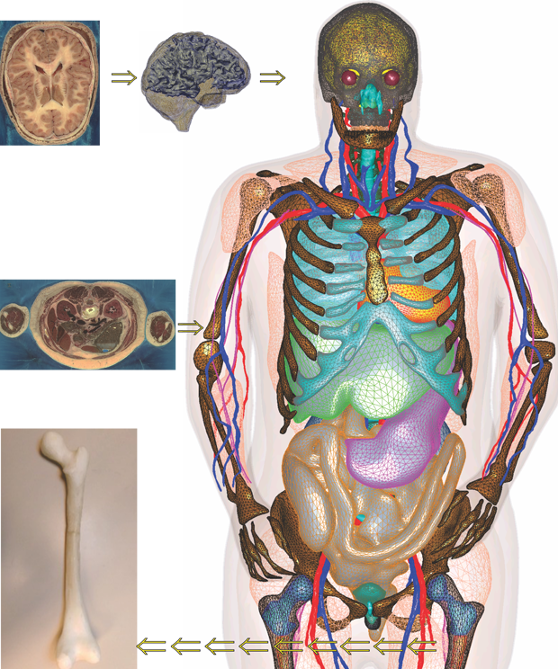
The VHP-Female model, which contains about 250 individual human bones, organs, tissues and structures (including additional parts such as hip implants), was based on over 5,000 high definition photographs of 0.33 mm thick anatomical slices in the axial plane of a deceased female patient, collected and archived as part of the Visible Human Project conducted by the National Library of Medicine (under the lead of Dr. Michael Ackerman). Each of these photos is defined by 24 bit color with a pixel resolution of 2048 by 1216 and each pixel is 0.33mm on a side. When taken together with the vertical spacing, these pixels create a 0.33mm3 voxel or volumetric domain of information. This offers an extremely rich and refined dataset from which to build a digitized model of a human being (Figure 1).
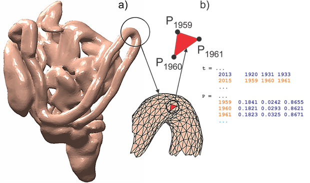
Each of the 250 components that are included in the model were painstakingly constructed through visual examination of the photographic data set and manually placing digital points or nodes at the boundaries of the object. For example, when constructing the model of the human heart, researchers combed through all the images of the VHP-Female thorax using image processing tools constructed in MATLAB and then carefully placed nodes around the edge of the heart, separating it from the remaining information in the photograph. This manual segmentation, when extended through all the slices, produces a three-dimensional ‘point cloud’, or set of coordinates in space that become the digital representation of the tissue. A surface surrounding the point cloud was then constructed by creating tiny triangular connecting points to each other, a process known as meshing. Each such surface or triangular mesh was constructed with reference to a global coordinate system that governs the entire model as shown in Figure 2. After the individual pieces of the model were created, each component was then processed to improve the quality of the triangular mesh – this dramatically increases the ability of any particular simulation software to then use the mesh. Each mesh was also tested with all other components to ensure that no mesh intersected with any other, which would create significant problems when executing simulations.
Once the various singular components of the model were completed, all were combined into a single project file that may then be used in commercial numerical software packages for solving complex problems such as electromagnetic wave propagation, energy absorption in human tissues, and the impact of medical device designs in their intended environments. But first, specific material properties, including electromagnetic permittivity and conductivity, must be assigned to each distinct mesh piece. These properties, which change with respect to operating frequency, are absolutely necessary to maintain accuracy in the simulation results. Data from a US Air Force study completed in 1996, which documents tissue properties from 10 Hz to 100 GHz, has been used in the VHP-Female model and includes all tissues ranging from the stomach to grey matter in the brain to the liver and lungs.
What Is It Used For?
Following proper material property assignment, the model is ready to support any number of biomedical and electrical engineering applications related to the human body. To date, a pool of approximately 100 users has employed the model to explore different applications and a number of diagnostic and therapeutic procedures that use focused electromagnetic stimuli to produce a response within the body. Doctors, engineers, and technicians are then able to ‘probe’ within the model at any desired location to assess the effectiveness of these procedures.
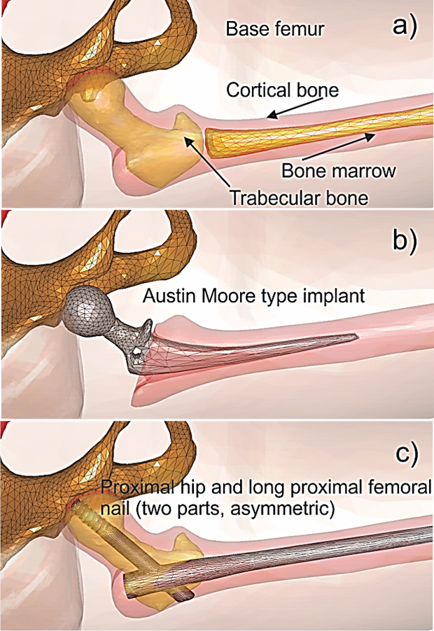
One application of interest is the production of localized heating during a magnetic resonance imaging (MRI) procedure due to the presence of metallic implants within the body. Three common metallic hip implants have been integrated into the model, shown in Figure 3, to gain perspective on this issue and assess any potential dangers to surrounding tissues that may exist. In a similar manner, the model may also be used to develop cancer ablation or detection techniques by inserting a cancerous tumor into a desired location and determining if a given procedure is effective.
Due to superior resolution in the head, the VHP-Female model also has been used to explore several non-invasive brain stimulation techniques used to treat ailments such as Parkinson’s disease, depression, Alzheimer’s disease, and other neurological disorders. One technique, known as Transcranial Direct Current Stimulation (tDCS) uses small amounts of electrical current introduced into the head through strategically placed electrodes to produced targeted stimulation of brain tissues. Transcranial Magnetic Stimulation (TMS), shown in Figure 4, employs a conducting loop placed above the head that passes very large currents through at relatively low frequencies. This current creates a magnetic field that painlessly passes into the body and generates eddy currents in the brain, which stimulate axon activation. However, the amount of resulting current within a given cranial tissue under stimulation with either of these techniques has, until now, been largely unknown.
The VHP-Female model has been used to provide accurate quantitative estimates when using these procedures, thereby influencing their use in clinical settings.
Can the VHP-Female Model Be Used With Other Programs?
The model is available in a variety of file types, including commonly used CAD formats (STL and SAT) as well as ANSYS High Frequency Structure Simulator (HFSS) and MAXWELL3D projects. The model is also available in MATLAB format, which enables its use for emerging technologies in customized numerical simulators and academic research codes [1]. All model components can be directly imported, viewed and manipulated either individually or as a whole within the base MATLAB environment without the use of additional toolboxes. This easily customizable format empowers researchers working in a variety of fields to explore the impact of their technologies and ideas on a realistic human body in a simulation environment, without the need for expensive instrumentation or extensive Institutional Review Board approvals. When used in MATLAB, the VHP-Female model also provides an ideal basis upon which to teach undergraduate and graduate level classes in electromagnetics, numerical methods, biomedical signal processing, and antenna design.
Next Steps for the VHP-Female Model
While the model in its current state has demonstrated tremendous utility in a variety of applications, development is far from complete. Work is currently underway to expand the definition of the model in several regions, including an expanded network of peripheral nerves and certain components of the endocrine system. The model has recently been thoroughly examined and evaluated by two leading electromagnetics software companies, the USA based ANSYS, Inc. and CST AG of Germany. In both instances, this process has led to licensing agreements, giving users of these software products exclusive access to the VHP-Female model in standardized and industry leading tools capable of simulating complex problems in electromagnetics and biomedical engineering.
Additional inspection of the model is being conducted by the Food and Drug Administration for its potential acceptance as a Medical Device Development Tool. Previous certification of the model has been granted by the IEEE International Committee on Electromagnetic Safety for determining spatial peak specific absorption rates in the human body by users of wireless communication devices.
In addition to the model expansion described above, a parallel effort is also underway to develop a detailed breathing sequence incorporating the fine internal detail provided by the VHP-Female model. This breathing sequence will model realistic external surface expansion experienced during respiration along with the appropriate displacement of surrounding interior bones and muscles involved. This development will enable FEM modeling of and investigations into the impact of dynamically varying spatial parameters and air volume in many applications, including physiological status sensor operation and RF channel modeling.
Reference
- S.N. Makarov, G.M. Noetscher, A. Nazarian, Low-Frequency Electromagnetic Modeling for Electrical and Biological Systems Using MATLAB, Wiley, June 2015.