New imaging techniques aid in patient diagnosis
In late February 2020, a time when severe acute respiratory syndrome coronavirus 2 (SARS-CoV-2, or COVID-19) still felt like an abstraction in the United States, New York City’s first infected patient was admitted to Mount Sinai Hospital’s emergency room. Working a few doors down was Sean Pinney, the Director of Advanced Heart Failure and Transplantation. Little did he know, but “that night was the beginning of hell,” he said.
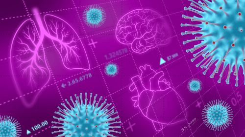
By March 1, the press caught wind of the infection. Panic ensued across the city, then cases began to surge. Pinney’s own first COVID-19 patient arrived days later, a man in his 40s, who suffered from severe pneumonia. Pinney prescribed the usual treatment—put him on a ventilator to keep oxygen flowing and keep his heart circulating blood. Days later, the man suffered sudden multiorgan failure and died. That was the moment Pinney realized that COVID-19 wasn’t just a bad pneumonia that patients would be able to just ride out on a ventilator. “There was a recognition that lurking behind all this must be some cardiovascular impact,” said Pinney (Figure 1).
Within a month, two-thirds of New York hospitals were filled to capacity with COVID-19 patients, and reports of a spectrum of cardiovascular ailments began to pop up—inflammation of the heart muscle known as myocarditis, heart attacks, and arrhythmia [1]. Approximately one quarter of hospitalized patients showed signs of injury to the heart and blood vessels. Many of those who did had needed ventilators when they were sick, many died. But then there were also young and active, otherwise healthy post-COVID heart patients who’d been barely symptomatic during their infections, but were showing up in Pinney’s office and others with similar heart and blood vessel damage [3]. Quite suddenly, Pinney and his peers around the world faced an urgent question: How did a respiratory virus cause this kind of damage, and how on earth would these doctors save their patients, especially as the novel coronavirus raced across the world?

At first, it just wasn’t clear how the virus could even affect the heart. The earliest reports of the virus described it mostly as a respiratory illness. But autopsy studies began to reveal a more complicated picture. In addition to causing damage to lungs, researchers found that in some cases the virus could also indirectly and directly impact the heart and related systems. Direct infection, called myocarditis, was less common—only 3% of COVID-19 autopsies show evidence of it—but in these cases, the virus invaded heart cells through their ACE2 receptors, the receptor gateways the coronavirus attaches to gain access to the inner cell, and which are present in a multitude of organs throughout the body including the heart, lung, gut smooth muscle, liver, kidney, and immune cells [2]. In myocarditis cases, the body’s ensuing immune response kills the virus, but often damages heart muscle in the process, sometimes leading to sudden cardiac arrest.
More often than not though, COVID-19’s cardiovascular impacts are more indirect, resulting from the residual effects of inflammation in the lining of blood vessels that can cause clotting or scarring, which constricts bloodflow. While clots can occur throughout the body—they have been seen in the lungs, kidneys, liver, and brain [2]—clots in the heart can be particularly deadly. Reduced bloodflow to the heart cuts off its oxygen supply, which leads to a heart attack and often sudden death.
Pinpointing potential cardiac problems before they start is critical for saving lives in COVID-19 patients, but knowing exactly what to look for isn’t straightforward, especially in those who appear otherwise healthy.
Long COVID
One of the most perplexing complications to occur in the course of this pandemic has been the phenomenon called “Long COVID”—what the National Institutes of Health refers to as “post-acute sequelae of COVID-19” [4]. This is a persistence of mostly COVID-like symptoms such as shortness of breath and fatigue, lasting at least four to eight weeks after the initial infection. Well over a year into the pandemic, there are those patients who have still not recovered from their illness, and others who will at times improve for a few months, only to be stricken anew by chest discomfort and brain fog [4]. When and whether these patients will ever fully recover is an open question.
In one of this year’s most common medical refrains, doctors and epidemiologists still don’t know exactly how prevalent long COVID is among patients, nor what the predisposing factors might be. Informal surveys so far indicated that anywhere between 2% to 20% of formerly infected individuals suffer from long-haul effects, and surprisingly, most of these patients are young and formerly healthy individuals—teenagers to middle-aged adults who had a light, if not asymptomatic, course of acute COVID [4].
The most common symptoms include heart palpitations, chest pain, and shortness of breath—but curiously, that doesn’t always line up with visible injury that a cardiologist can see. “I had a feeling when I first started seeing the long-haul patients that I was going to see a lot of abnormal cardiac MRIs[magnetic resonance images]” said Pinney. Instead, his standard imaging and echocardiogram data rarely show evidence of inflammation or scarring. And yet, “I have seen people with legitimate chest pain, and I can’t explain where it’s coming from,” he said. Numerous studies using traditional screening methods have reported similar symptoms with no clear evidence of heart damage [5].
One reason for Pinney’s conundrum could be that the damage his patients experience is difficult to find using traditional measures. Valentina Puntmann, a cardiologist and head of the Experimental and Translational Cardiovascular Imaging Department at the University of Frankfurt in Germany (Figure 2), has been refining and developing advanced techniques to image inflammation throughout the body, particularly in the heart, and discovered a very different pattern from Pinney.
When the first pandemic wave hit Germany in the summer of 2020, Puntmann and her colleagues distributed flyers advertising free cardiac MRIs at the University Hospital. At the time, it was forbidden to image anyone in the acute stage of infection, so all recruits would have already recovered from the virus. “We still thought of COVID as a lung infection then, so we didn’t think we’d find anything,” she said.

Puntmann uses a new type of cardiac MRI technique called T1 and T2 mapping to detect heart inflammation, a tool that is far more sensitive than the typical cardiac MRI. A typical cardiac MRI detects differences between tissues across the heart, which is useful for something like a heart attack where one portion of the heart loses bloodflow and oxygen and dies. In the case of inflammation, however, the entire heart may be inflamed, as well as the area surrounding it, and so absent discrete differences within the organ, problems can be more easily missed. T1 and T2 mapping, however, analyzes the muscular contractions of the heart itself, and can measure whether or not it is struggling to pump blood, which would be indicative of an inflamed muscle [3].
Puntmann performed T1 and T2 cardiac MRIs on 100 formerly infected people, and incredibly, a solid three-fifths of them showed signs of myocarditis. The results were quickly published in JAMA Cardiology in July 2020 [3]. Later that year, another study reported similar results in a smaller cohort of 26 Ohio State athletes who had recovered from mild COVID-19 [6]. “People don’t understand why we found so many things and others didn’t,” said Puntmann. “I think this is because most people don’t understand that there are differences between different methodologies.”
Imaging as the answer
Pre-pandemic, undiagnosed myocarditis was the leading cause of death in otherwise healthy young athletes. The heightened activity puts additional strain on a struggling heart. It’s a nightmarish situation, but now in the wake of the coronavirus pandemic, a cohort of young athletes have been placed into that exact vulnerability. Even without symptoms, exercise within the first three months of an infection can lead to long COVID, said Puntmann. She advises patients to keep their heart rates below 100 during that time period.
The current risk scenario makes it critical for doctors to create rapid myocarditis diagnostics. Absent Puntmann’s experimental T1 and T2 MRI technique, the gold standard for diagnosing myocarditis involves lengthy blood tests or a cardiac biopsy, an invasive procedure where a piece of heart muscle is removed through one of the body’s larger veins, usually located in the neck or thigh.
Given that the rates of long COVID range from 2% to 20% of potentially all infected individuals, at least some of them young and otherwise healthy, blood tests and biopsies become wholly impractical. Imaging takes less time, but with the sheer number of patients, a doctor’s time could be better spent treating patients than diagnosing them.

Jasjit Suri (Figure 3), a biomedical engineer and medical imaging specialist, thinks that image-based artificial intelligence (AI) can be used to characterize the tissues of a COVID-19 patient and classify the severity of their infection, cutting down on the time it takes to identify an urgent cardiovascular problem before it’s too late. A cardiac MRI, once taken, spits out a complex three-dimensional image that must be viewed from multiple angles and in many different ways to make a diagnosis. AI can be programmed to automate this process, reading different features within an image to flag potential problems in patients on the cusp of a dangerous, potentially fatal episode. It would reduce the amount of time from test to treatment [7].
Suri is the CEO of AtheroPoint, a medical imaging technology company that has developed software that does exactly that for a variety of ailments in the liver, thyroid, heart, and other organs. Before the pandemic, it had already been deployed in hospitals around the world, making the diagnostic process for numerous ailments faster and more efficient. “We are able to quickly predict the risk and severity of blocked arteries and blood vessels,” using the software, said Suri. By training the software to identify potential COVID-induced blockages in the heart, he thinks the software can quickly predict problems before they start. But, “the gold standard for diagnosis is always human,” he said, “this is merely a tool to save time.” Plus, the software’s use in diagnosing COVID-related cardiovascular issues is, for now, just an idea [7]. Suri hopes to test AtheroPoint’s software in hospitals soon, but it will take some time before it’s ready to be deployed in earnest.
Treating broken hearts
There is one fundamental problem with AI and T1/T2 cardiac imaging—you need access to the technology, and the money to pay for it. With hundreds of millions infected, many in regions with limited access to healthcare, it would be helpful to have approved pharmaceutical treatments that are readily and easily dispensed. We already have cheap drugs, betablockers, for cardiac inflammation, said Puntmann, but studies have not yet been completed to prove their efficacy in the context of COVID-19. “We need to produce much more of a scalable approach to treating this disease similar to how we treat the sore throat,” said Puntmann, citing the known safety of betablocker drugs, which she believes would only need to be used temporarily until a patient’s myocarditis subsides.
“I’m really afraid this is not going to go away,” said Pinney. Pandemics have been known to circulate in the human population for thousands of years—take smallpox, which dates back to Ancient Egypt [8]. “What if it keeps moving around and around the world? We don’t know how long we will be protected from the vaccine,” he said, “I really don’t want to live through another surge. I think all of us are just exhausted by COVID.”
References
- M. Chilazi, E. Y. Duffy, A. Thakkar, and E. D. Michos, “COVID and cardiovascular disease: What we know in 2021,’’ Current Atherosclerosis Rep., vol. 23, no. 7, p. 37, 2021.
- G. Giustino et al., “Coronavirus and cardiovascular disease, myocardial injury, and arrhythmia: JACC focus seminar,’’ J. Amer. College Cardiol., vol. 76, no. 17, pp. 2011–2023, 2020.
- V. O. Puntmann et al., “Outcomes of cardiovascular magnetic resonance imaging in patients recently recovered from coronavirus disease 2019 (COVID-19),’’ JAMA Cardiol., vol. 5, no. 11, pp. 1265–1273, 2020.
- Z. Al-Aly, Y. Xie, and B. Bowe, “High-dimensional characterization of post-acute sequelae of COVID-19,’’ Nature, vol. 594, pp. 259–264, Apr. 2021.
- N. Moulson et al., “SARS-CoV-2 cardiac involvement in young competitive athletes,’’ Circulation, vol. 144, no. 4, pp. 256–266, 2021.
- S. Rajpal et al., “Cardiovascular magnetic resonance findings in competitive athletes recovering from COVID-19 infection,’’ JAMA Cardiol., vol. 6, no. 1, pp. 116–118, 2021.
- J. S. Suri et al., “COVID-19 pathways for brain and heart injury in comorbidity patients: A role of medical imaging and artificial intelligence-based COVID severity classification: A review,’’ Comput. Biol. Med.,
vol. 124, pp. 103960–103960, Aug. 2020. - R. B. Kennedy, I. Ovsyannikova, and G. A. Poland, “Smallpox vaccines for biodefense,’’ Vaccine, vol. 27, pp. D73–D79, Nov. 2009.