
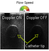
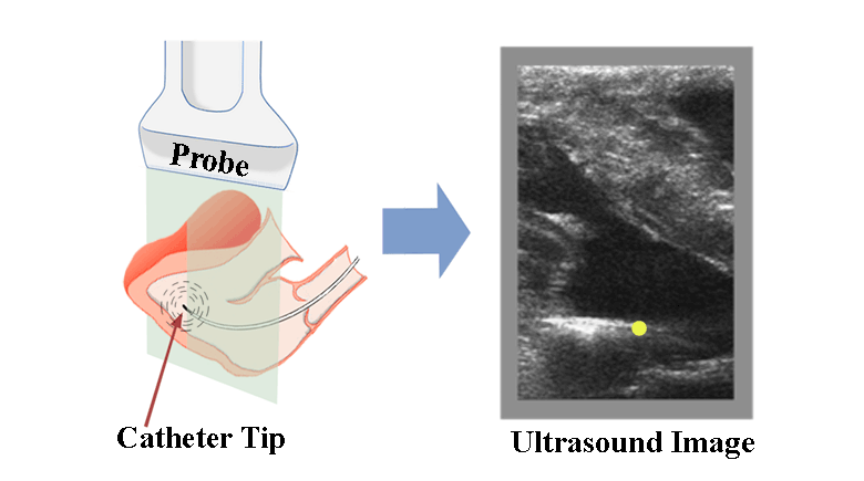
Ultrasound-guided navigation of intracardiac catheters during minimally invasive procedures can be challenging due to the inability of B-mode ultrasound to distinguish the distal end of catheter from its body and the surrounding tissue. To better visualize the distal tip and guide the catheter, a prototype acoustically active catheter with a miniature vibrating piezoelectric crystal is used. The vibrating crystal interacts with the imaging ultrasound field creating symmetric Doppler shifts at frequencies equal to the crystal vibration frequency along with higher harmonics. A novel symmetric Doppler shift detection algorithm has been proposed to leverage this unique physical phenomenon for identification of the catheter tip. Using Doppler ensemble, the algorithm identifies the image pixels where both the positive and negative Doppler shift frequencies exist that pass a minimum signal to noise threshold. The identified points can then be overlaid with a unique color (Yellow) on a B-mode scan with or without a color Doppler overlay of blood flow. The technique was optimized to avoid interference from blood flow and tested in real time at a frame rate varying between 22 to 50 Hz, depending upon the size of the field of view. The catheter tip can be localized precisely in all three axes with localization distance equal to 1.8 mm in axial and lateral direction and 2.4mm in elevational direction. The technique is able to detect the distal tip even when the signal from the blood is on par in amplitude with the vibrating piezoelectric signal. The technique can be easily ported to clinical ultrasound machines with minor changes to the Doppler processing software facilitating unambiguous and distinct real-time visualization of the distal tip of intracardiac catheters.