Hardik J. Pandya, Kihan Park, Wenjin Chen, Lauri A. Goodell, David J. Foran, and Jaydev P. Desai, Brigham and Women’s Hospital – Harvard Medical School, University of Maryland, Rutgers Cancer Institute of New Jersey, Rutgers Robert Wood Johnson Medical School, Rutgers USA.

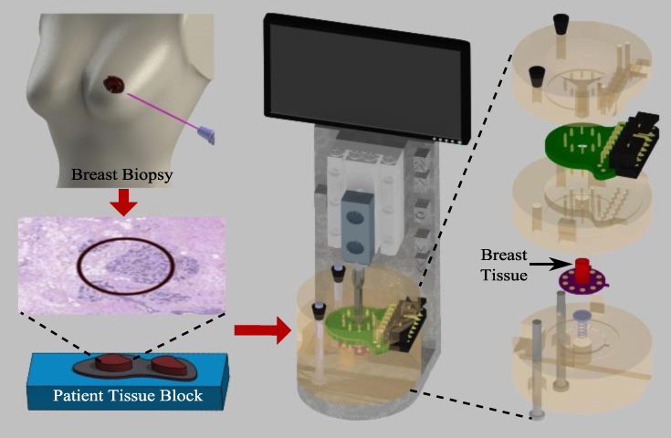
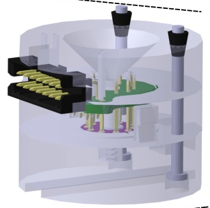
Breast cancer is characterized by progressive modifications of the microenvironment of the mammary tissue; the innate, tightly-controlled, mesenchymal-stroma microenvironment undergoes biochemical changes and physical restructuring to become tumor-permissive. A collagen-rich desmoplastic stroma is one of these distinctive developments and it causes changes in tissue elasticity. Being able to monitor these tissue changes can be critical to improving clinicians’ diagnostic capabilities and developing early intervention strategies. Needle biopsy of the breast is a gold standard to diagnose breast cancer. Microscale devices can operate on specimens sized similar to biopsy samples and provide a pathway to gain further insight into mechanical, electrical, histological, and immunohistochemical profile changes in breast tissue, from onset through disease progression, and can be used to demarcate the specific stages of cancer in epithelial and stromal regions, providing quantitative indicators facilitating the diagnosis of breast cancer. We have used an interdisciplinary approach of merging microengineering (Micro-Electro-Mechanical-Systems) and additive manufacturing (3D printing) to develop a portable cancer diagnostic tool which can be used to study the underlying architectural changes (Electro-Thermo-Mechanical) that occur during the progression of breast cancer at the micro level. The tool is capable of delineating normal and cancerous breast tissues and can potentially be used to study other tissue related cancers.
Keywords: Biomedical microelectromechanical systems, breast cancer diagnosis, tissue engineering, Biomedical measurement, Biosensors, Breast tissue

