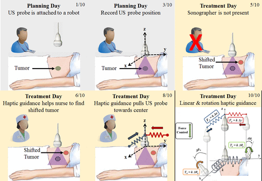
Image-guided radiation therapy (IGRT) involves two main steps: (1) planning/simulation using computerized tomography (CT) imaging and (2) fractionated treatment delivery using a linear accelerator (LINAC). These are performed on different days and in different rooms; thus it is necessary to reproduce the patient position so that the delivered dose closely matches the planned dose. This can be challenging for soft-tissue targets, especially abdominal organs, because they are difficult to visualize in the cone-beam CT images acquired by the LINAC in the treatment room. Ultrasound imaging can overcome these problems, but an ultrasonographer would be required to locate the target organ and the probe contact force could deform the tissue, thereby causing discrepancies with respect to the treatment plan. We introduce a cooperatively-controlled robot system for IGRT in which a clinician and robot share control of a 3D ultrasound (US) probe. To compensate for soft tissue deformations created by the probe, we present a novel workflow where the robot holds the US probe (or x-ray compatible model probe) on the patient during acquisition of the planning CT image, thereby ensuring that planning is performed on the deformed anatomy. An ultrasonographer is still required on the planning day to acquire the reference US image, but the robot system enables the radiation therapist to produce consistent soft tissue deformation between simulation and treatment days. The system considers the robot position, contact force, and reference US image recorded during simulation and introduces constraints (virtual fixtures) to guide the radiation therapist. The proposed clinical workflow is validated by an in-vivo canine study, which suggests that the proposed cooperative control method with soft virtual fixtures (implemented as virtual springs) can improve the reproducibility of patient setup and soft tissue deformation during the fractionated radiation treatment. Further information is available at https://smarts.lcsr.jhu.edu/research.

