
Optical microscopy is an indispensable technique for investigating neuronal morphology and dynamics due to the high spatial and temporal resolution granted by light. However, neuronal morphology and structural changes occur on scales beyond the optical resolution of canonical microscopy technologies. Super-resolution light microscopy (SRLM), such as photon localization microscopy, stimulated emission depletion (STED) microscopy, and structured illumination microscopy (SIM) can overcome Abbe’s diffraction limit and increase optical resolution. These SRLM technologies have proven valuable in biological studies, especially in evaluating neuronal dynamics and the morphology-function relationship of neurons. Unfortunately, SRLM generally relies on the incident and emitted photon structures remaining intact during imaging. This limitation constrains the application of SRLM techniques in scattering tissues, reducing their imaging speed or imaging depth, or both. As a consequently, SRLM is confined primarily to investigations of cell cultures, as well as acute or organotypic brain slices.
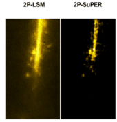
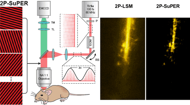
One method to improving SRLM imaging depth while maintaining sufficient imaging speed involves combining SIM with two photon laser scanning microscopy (2P-LSM). 2P-LSM increases imaging depth by using a low-scattering near infrared (NIR) excitation laser beam. In the case of patterned illumination, 2P excitation can maintain illumination patterns deeper into scattering tissue when compared to single photon illumination. Here, we apply our technique, resonant two-photon Super-resolution Patterned Excitation Reconstruction (2P-SuPER) microscopy, to improve SRLM imaging depth and speed in scattering tissues. 2P-SuPER combines fast resonant scanning with the improved imaging depth of 2P-microscopy imaging rates of 3.5 Hz at depths past 100 μm in living scattering tissue while maintaining the two-fold resolution enhancement of SIM. We demonstrate the capability of our technique by imaging eGFP in the cytosol of neurons in acute brain slices and in the neocortex of anesthetized mice.