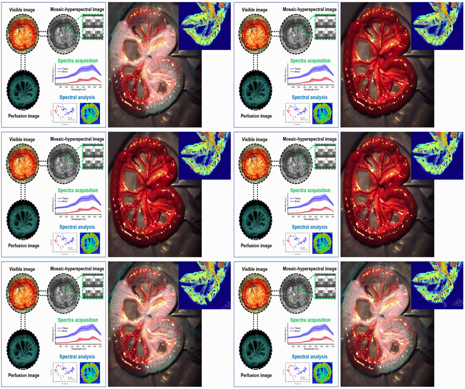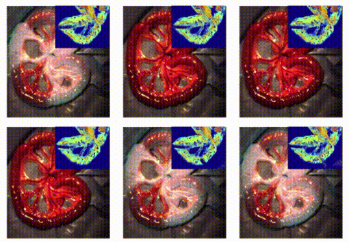
Real-time monitoring of patient status is required to increase the likelihood of successful blood supply and reduce the risk of organ damage in precise surgical operations such as tissue resection and transplantation. This study introduced a multimodal surgical guidance system that simultaneously monitors blood vessel perfusion, oxygen saturation, thrombosis, and tissue recovery using a combination of visible imaging, mosaic filter-based hyperspectral imaging (HSI), and laser speckle contrast imaging (LSCI). This convergence technology does not require the use of a label or contrast agent, or delay for drug injection or diffusion. Blood vessels in the small intestine of rats were clamped to create areas of restricted blood flow. The subsequent tissue damage and regeneration were monitored immediately after surgery and were compared to the control region. Blood supply was confirmed by measuring vessel perfusion using LSCI to monitor the biological activity and survival of organ tissues. Meanwhile, blood oxygen saturation was monitored using HSI in the near-infrared region. Principal component analysis (PCA) across wavelengths was used to optimize the HSI spectral combination for hemodynamic biomarker visualization. HSI and LSCI were used to identify thrombus generation and tissue recovery. The prognosis for recovery was also determined based on the blood supply to the organ using integrating tissue imaging information from visible imaging, LSCI perfusion imaging, and HSI. This convergence of complementary imaging devices can operate in real time to determine blood supply and tissue prognosis during surgery. Based on these technologies, the risks to patients in the operating room can be minimized.

