
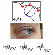
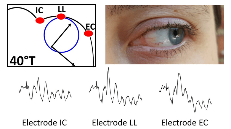
To objectively asses retinal function, ophthalmologists use the electroretinogram (ERG): the electrical signal produced by the retina in response to a flash of light. However, because it uses a single electrode and a full-field stimulus, only a global signal is obtained, thus preventing the localization of retinal dysfunction or damage. To overcome this limitation, this work aims at establishing a new recording protocol where the number of recording electrodes is increased without encumbering the limited area around the eye. To achieve this goal, we placed 3 real electrodes around the eye, and combined this with 11 gaze positions thus generating 33 virtual electrodes which permitted the recording of 33 different derivations of the ERG signal. We named this novel ERG recording protocol the multi-angular ERG (or maERG). With these varying recordings and using a realistic electrophysiological and anatomical eye model, we reconstructed the retinal activity which would have produced the 33 maERG signals by solving the inverse problem.
To assess feasibility and accuracy of our technique, we simulated diseased retinas with central or peripheral scotomas of varying sizes, as well as varying levels of noise added to our simulated signals. With these simulated signals, two inverse problem-solving methods were compared in this study, where we found that the low-resolution brain electromagnetic tomography algorithm (LORETA) performed better at correctly reconstructing the scotoma than the Minimum Norm Estimate (MNE) algorithm. With LORETA, we established a signal to noise ratio limit of 50dB was further needed. Thus, we demonstrate here that it is possible to accurately reconstruct the retina’s functional map, given a good enough signal to noise ratio and our solution to the electromagnetic inverse problem is followed. This novel functional retinal imaging technique will permit an optimized clinical monitoring of patients affected with retinopathies, by localizing and detecting retinal malfunction earlier.