There is often more than meets the eye when assessing tissue health. Important tissue health biomarkers may not be visually apparent at surface level. Optical imaging systems assess tissue composition and structure using non-invasive light-based techniques by analysing the molecular composition of the absorbing and scattering agents. Diffuse optical spectroscopic imaging (DOSI) is one such biophotonic, or light-based, technology capable of assessing deep sub-surface tissue properties. The probe is traditionally placed at a specific location, and a point measurement of the tissue composition is determined through frequency modulated light through a source-detector pairing. Clinical trials have shown tremendous promise for safe (no ionizing radiation) breast cancer assessment, however producing a full spatial tissue map is traditionally laborious, requiring many manual point-based measurements, posing a significant barrier to clinical adoption.
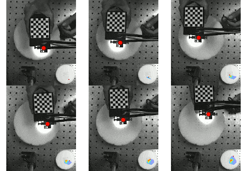
Here, we propose a camera-based solution for collecting whole area tissue optical property maps in real-time. A single calibrated overhead camera was used to track the DOSI probe in 2D frames, and a fiducial target (in this case, a checkerboard) was printed on the top probe face. By incorporating an a priori model of the target geometry, and knowing the calibrated camera properties, the 3D position and orientation of the probe could be determined from these 2D frames, providing its measurement location on the tissue surface. This 3D location was concurrently processed with each optical measurement, and a spatial optical property map was computed through 2D interpolation. The system exhibited sub-millimeter system accuracy within the camera’s field of view across different probe speeds, enabling fast hand-guided scans. This system provides a valuable tool for real-time non-invasive tissue health and cancer screening, and enables longitudinal disease progression assessment through unstructured probe-based optical tissue assessment.

