Peter A. Galie, Fitzroy Byfield, Christopher S. Chen, J. Yasha Kresh, Paul A. Janmey, University of Pennsylvania, Boston University, Drexel University, Volume 62, Issue 2, Page:438-442

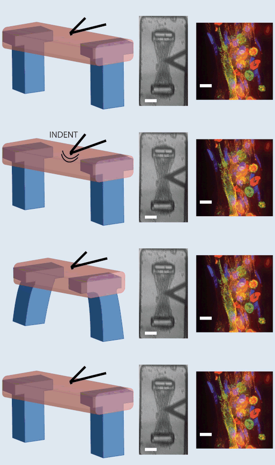
In recent years, microscale tissue gauges have provided a platform to study several aspects of cardiac electrophysiology in a three-dimensional, mechanically controlled environment. Combining this system with an atomic force microscope, we have demonstrated a method for studying the mechano-electric coupling of in vitro models of cardiac tissue. In contrast to previous studies performed on 2D surfaces coated with extracellular matrix proteins, which is not as representative of the in vivo myocardium, the current setup allows for the study of mechano-electric coupling in a three-dimensional geometry. The tissue analogs consist of fibroblasts and cardiomyocytes isolated from neonatal rat ventricles and then embedded within composite matrices of collagen and fibrin. Cyclic, small indentations (< 3 microns) into the approximately 60 micron-thick constructs with the microcantilever of an AFM probe induce a coordinated contraction in the absence of any electrical fields. The contractions of the constructs occur at an approximately 1 Hz regardless of indentation depth or frequency. However, increasing indentation frequency does increase the time to peak of the contraction cycle, and the constructs require continuous indentations to maintain the beating response. Overall, mechanical energy delivered using percussive stimulation was shown to affect cardiac electrophysiology through mechano-electric coupling and feedback. This result has several implications for mechanically-induced arrhythmia generation, and other disorders that arise from disrupted mechano-electric coupling in the heart.
Keywords: Mechano-electric Coupling, Cardiac Electrophysiology, Mechanical Stimulation, Mechanosensitivity

