
Anthony G. Christodoulou, T. Kevin Hitchens, Yijen L. Wu, Chien Ho, and Zhi-Pei Liang, University of Illinois at Urbana-Champaign and Carnegie Mellon University, Volume 61, Issue 9, Pages: 2451-2457

Cardiac magnetic resonance imaging (MRI) is relatively slow compared to other imaging modalities, Sparse sampling methods have emerged as effective tools to accelerate cardiac magnetic resonance imaging (MRI), including low-rank model-based methods. Low-rank model-based methods commonly assume an explicit temporal subspace in order to reconstruct images from highly undersampled (k,t)-space data; this temporal subspace is most commonly estimated from navigator data collected after every other RF pulse using Cartesian trajectories. However, this navigation strategy is temporally inefficient and can miss temporal information, as Cartesian trajectories have a null space preventing them from detecting perpendicular translation. Despite these issues, little work has been published on navigation strategies to improve subspace estimation before now.
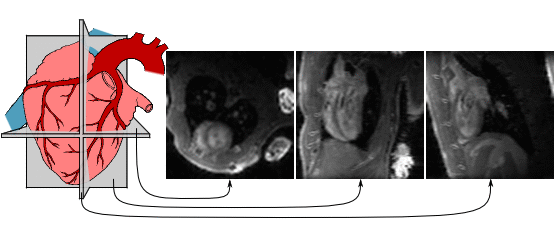
In this paper, we propose strategies to improve subspace accuracy and increase imaging speed. We investigate the use of non-Cartesian k-space navigator trajectories such as spirals, cones, and a novel “music note” trajectory to replace the ubiquitous Cartesian navigator trajectories, increasing subspace accuracy. We also introduce efficient “self-navigated” pulse sequences that collect both navigator and imaging data after every RF pulse, further accelerating cardiac MRI. We have evaluated these navigation strategies through analysis of phantom images, and we have demonstrated their use for in vivo cardiac imaging of rats and mice without the use of ECG or respiratory gating. The proposed methods achieved 3-D imaging of wall motion, first-pass myocardial perfusion, and late gadolinium enhancement in rats at 74 frames/s, as well as 2-D imaging of wall motion in mice at 97 frames/s.
Keywords: Cardiovascular magnetic resonance imaging (MRI), low-rank imaging, MRI pulse sequences, partial separability (PS), subspace estimation.