
The integration of the multi-channel measurement capabilities of near-infrared spectroscopy (NIRS), electrocorticography (ECoG), and negative temperature coefficient (NTC) thermistor sensors into a single device, having a compact profile similar to that of the subdural strip electrodes commonly used in neurosurgery, can provide beneficial information on various aspects of brain cortical activity and prove to be a powerful medical modality for pre-, intra-, and post-operative diagnoses.
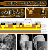
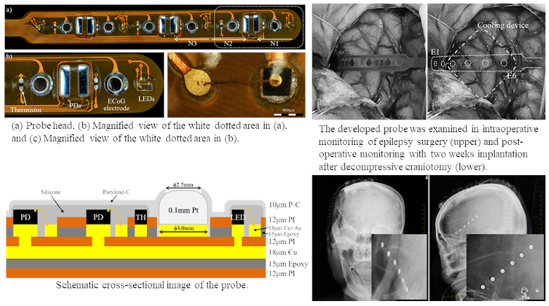
This paper describes the development of a flexible multi-modal multi-channel probe for the simultaneous measurement of the NIRS, ECoG, and surficial temperature obtained from the cerebral cortex. Photoelectric bare chips for NIRS channels, miniature temperature-coefficient thermistors for measuring localized temperature variation, and 3-mm-diameter platinum plates for ECoG recording were assembled on a polyimide-based flexible printed circuit to create six channels for each modality within a total thickness of 0.7 mm. A conformal coating of Parylene-C was applied on all the channels except the ECoG electrodes to make the probe surface biocompatible.
As a first-in-human study, the simultaneous measurement capability of the multi-modality probe, with sufficient signal-to-noise (S/N) ratio and accuracy, to observe pathological neural activities in subjects during surgery and post-operative monitoring was examined with IRB approval. The measured signals during epilepsy surgery with the focal brain cooling procedure showed suppression of epileptogenic activity in ECoG, the dilution of oxy-hemoglobin concentration in NIRS, and the temperature degradation in the thermistors during the cooling period. The placement of the developed probe on the ipsilateral frontal lobe surface during decompressive craniotomy for 14 days recorded the neuropathological phenomenon of cortical spreading depolarization in all modalities, with no complication due to two-week implantation.
The simultaneous and accurate multi-channel recording of electrical, hemodynamic, and thermographic cortical activities in a single device small enough for subdural implantation is likely to have major implications in neurosurgery and neuroscience.